

Аннотация
Rhamnus alaternus L. is a Rhamnaceae shrub and a popular traditional medicine in Algeria. The present research objective was to investigate the antioxidant, genotoxic, and antigenotoxic properties of R. alaternus methanolic leaf extract.Antiradical scavenging activity was tested by α, α-diphenyl-β-picrylhydrazyl free radical scavenging and β-carotene bleaching method. DNA damage and repair were measured by the Allium cepa test with sodium azide as a mutagenic agent. Mitotic index and chromosomal aberrations were calculated by microscopy of meristem roots stained with 2% carmine acetic.
The methanolic extract of R. alaternus leaves inhibited the free radical DPPH (IC50 = 0.74 ± 0.30 mg/mL) and prevented the oxidation of β-carotene (50.71 ± 4.17%). The root phenotyping showed that sodium azide changed their color and shape, decreased their stiffness, and significantly reduced their length. The roots treated with both R. alaternus leaf extract and sodium azide demonstrated a better root growth. The roots treated with the methanolic extract were much longer than the control roots (P < 0.001). The microscopy images of root meristem treated with the sodium azide mitodepressant agent showed significant chromosomal aberrations, which indicated a disruption of the cell cycle.
The R. alaternus leaf extract appeared to have a beneficial effect on cytotoxicity. The antioxidant properties of R. alaternus L. makes this plant an excellent genoportector.
Ключевые слова
Rhamnus alaternus L., antioxidant activity, Allium cepa, chromosomal aberrations, antigenotoxicity, mitotic indexВВЕДЕНИЕ
Radical oxygen species lead to cell damage, which can induce genetic instability responsible for many pathological processes. This damage can be repaired by some natural compounds, e.g. radical scavengers and powerful protective antioxidants [1]. Kitagishi et al. proved that medicinal herbs could one day become a promising therapeutic means of cancer therapy [2]. According to Dayani et al., antioxidant, antiinflammatory, and anti-apoptotic properties of plants and their derivatives make them good radioprotectors against the mutagenic action of X-rays [3].
Phytotherapy relies on medicinal plants and their active compounds. Rhamnus alaternus L. (Rhamnaceae family), also called imlilesse or safir in the North of Algeria, is well known for its biological properties [4]. Zeouk and Bakheti reported that a decoction of the aerial part of the R. alatrenus leaves and branches has been widely used in traditional medicine to lower blood pressure and treat hepatitis, icterus, musculoskeletal disorders, and gastrointestinal diseases. They also serve as a cataplasm for skin infections [5].
Previous findings proved that R. alaternus extracts possess potential antioxidant, cytotoxic antimutagenic, antigenotoxic, and antimicrobial activities [5–8]. In their bibliographic review, Nekkaa et al. focused on the phytochemical and pharmacological properties of R. alaternus [4]. Its leaf extracts were rich in flavonoids, tannins, and anthocyanins, which explains their potential antigenotoxic and antimutagenic activity.
Bhouri et al. isolated kaempferol 3-O-b-isorhamninoside and rhamnocitrin 3-O-b-isor-hamninoside from R. alternus leaves [9]. These flavonoids are effective free radical scavengers and potent antigenotoxics. However, they can induce apoptosis in human lymphoblastoid cells by the extrinsic apoptotic mechanism including DNA fragmentation, PARP cleavage, and active caspase-3 and caspase-8 [10]. Oligomer flavonoid extract from R. alaternus leaves proved to have a good potential for alternative antimelanoma therapies [11].
Although some plant remedies have welldocumented protective effects and alleviate many diseases, cytotoxicity studies are very important for developing new drugs. Gadouche et al. described the toxic effect of Aristolochia longa L. and Calycotome spinosa L. on the blood cells and concluded that it should be studied on cancer cells [12]. Natural antioxidants can even protect human organism against the cytotoxic and mutagenic effects of xenobiotics.
In this research, we analyzed the genotoxic and DNA damage protecting activity of R. alaternus leaf extract by using the Allium cepa assay with azide sodium as a mutagen agent.
ОБЪЕКТЫ И МЕТОДЫ ИССЛЕДОВАНИЯ
Plant material. The research featured Rhamnus alaternus eu-alaternus L., a subspecies of Rhamnus alaternus L. The samples were collected in the Bissa forest located in the north of the Chlef province (Algeria). This species of Algerian flora was identified by Dr. Belhacine, a botanist from the Chlef University [13]. The R. alaternus leaves were dried in the dark for 10 days. After that, they were ground into a fine powder and kept in an airtight container, and 10 g of the dry powder was macerated in 100 mL of petroleum ether for 24 h with stirring. The mix was filtered on Whatman No. 1 paper. The maceration included 100 mL of methanol. After filtration, the marc was evaporated in a rotary evaporator at 39°C. The extract obtained was stored at 4°C until use [14].
Quantitative analysis and antioxidant activity. The polyphenols were assayed according to the method developed by Raafat and Samy [15]. The amount of total polyphenols was determined spectrophotometrically using the Folin- Ciocalteu reagent and deduced from a calibration curve established with gallic acid (0–1 mg/mL). The results were expressed in mg of gallic acid equivalent per g of dry matter (mg GAE/g of dry matter). The mix included 250 μL of Folin Ciocalteu’s phenol reagent, 50 μL of each concentration prepared from stock solution, and 500 μL of 20% Na2CO3 aqueous solution. After vortexing, the solution was adjusted with 5 mL of distilled water. After 30 min of incubation, the absorbance was measured at 765 nm. The same procedure was carried out with the extract obtained from R. alaternus leaves.
The flavonoid content was assayed according to the method developed by Hmid et al. [16]. After 1 mL of extract was added to 1 mL of 2% aluminum chloride, the absorbance was determined at 430 nm after 10 min of incubation. Quercetin served as calibration curve standard and was established from the concentration of 40 μg/mL of stock solution. Total flavonoids content in the extract was expressed as mg quercetin equivalents per g of sample (mg EQ/g of dry matter).
The DPPH assay followed the method described by Burits and Bucar [17]. The R. alaternus extract had the following concentrations: 0.2, 0.4, 1.0, and 2.0 mg/mL. We mixed 50 μL of each concentration with 5 mL of 0.004% DPPH. The absorbance was measured at 517 nm after 30 min of incubation. The results were compared to ascorbic acid, which was used as standard antioxidant and handled under the same conditions. The percentage of inhibition and IC50 were calculated according to Sharififar et al. [18]. The percentage inhibition was calculated using the following equation:
where OD is optical density.
IC50 is the concentration of extract required for 50% inhibition of DPPH. It was calculated using a linear regression analysis.
The β-carotene-linoleic acid assay was performed according to the method described by Kartal et al. [19]. The emulsion included 0.5 mg of β-carotene, 1 mL of chloroform, 25 μL of linoleic acid, and 200 mg of tween 40. The chloroform was eliminated in a rotary evaporator under vacuum, and 100 mL of distilled oxygen-saturated water was added to the emulsion. Subsequently, 350 μL of the extract at a concentration of 2 mg/mL was mixed with 2.5 mL of the emulsion. After 48 h of incubation, the absorbance was registered at 490 nm and compared with that obtained with butylohydroxytoluene (BHT), which served as a standard antioxidant and was prepared under the same conditions. The inhibition percentage of bleaching (I, %) was measured for each assay using the following equation:
![]()
Allium cepa assay. The A. cepa assay was performed according to Tedesco and Laughinghouse with some modifications [20]. The onion bulbs were kept in a culture medium that included 60 mg/L of CaSO4, 60 mg/L of MgSO4, 96 mg/L of NaHCO3, and 4 mg/L of KCl. They were incubated at 25°C for 72 h until the roots reached 2 cm. Seven onion bulbs were utilized for each treatment as follows: Sample 1: culture medium + distilled water; Sample 2: culture medium + sodium azide (50 mg/mL); Sample 3: culture medium + sodium azide (100 mg/mL); Sample 4: culture medium + methanolic extract (50 mg/mL); Sample 5: culture medium + methanolic extract (100 mg/mL); Sample 6: culture medium + sodium azide (100 mg/mL) + methanolic extract (50 mg/mL); Sample 7: culture medium + sodium azide (100 mg/mL) + methanolic extract (100 mg/mL).
The effect of the different treatments on the growth (cm) of the A. cepa roots was measured at different time intervals: 0, 24, 48, and 72 h. In parallel, the roots were tested for color, shape, and stiffness. After each time interval, the roots were collected for microscopic observation of the meristem cells and stored in 70% ethanol for later use. The roots were fixed in acetic acid and ethanol solution (1:3) for 24 h. After triple rinsing with distilled water, the roots were hydrolyzed with HCl (1N) and incubated in a hot water bath at 60°C for 10 min. After the hydrolysis, the roots were rinsed once again in distilled water and stained with 2% acetic carmine in a hot water bath at 60°C for 10 min. After incubation, the terminal meristem cells of the colored roots were cut with a scalpel under a binocular magnifier. The meristem regions were crushed manually between blade and coverslip to visualize the chromosomes and the different stages of cell division. Meristem cells were counted for each sample and tested for normal or abnormal cell division in search for mutations. The mitotic index and the rate of aberrant cells of each bulb were calculated by the following formula [21]:
![]()
![]()
Statistical analysis. The experimental data were analyzed using the ExcelSTAT software. The research also included the ANOVA variance analysis, followed by the Tukey’s test. The statistically highly significant value was P < 0.001.
РЕЗУЛЬТАТЫ И ИХ ОБСУЖДЕНИЕ
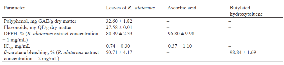
The total phenol content in Rhamnus alaternus L. leaves was 32.6 ± 1.82 mg GAE/g DM, and the total flavonoid content was 27.58 ± 0.01 mg EQ/g DM. The methanolic extract of R. alaternus leaves demonstrated a moderate efficiency against free radicals emitted by linoleic acid (50.71 ± 4.17%). Its capacity to beat free radical of DPPH (I% = 80.39 ± 2.33%, IC50 = 0.74 ± 0.30 mg/mL) was close to that of ascorbic acid, i.e. 96.80 ± 9.98% with IC50 = 0.37 ± 1.10 mg/mL (Table 1).
Plants are excellent indicators of the cytotoxic, cytogenetic, and mutagenic effects of environmental chemicals. They can serve as an alternative for detecting possible genetic damage in cells [22]. Genotoxicity studies were carried out by the Allium cepa assay. This method provides a convenient in vivo model to evaluate cell cycle alterations induced by mutagens [20].
The A. cepa roots treated with distilled water had no morphological change: the growth rate was good, the color was whitish, and the roots were rigid and bulky. However, the roots treated with sodium azide (50 and 100 mg/mL) changed the color and shape of the roots, as well as reduced their rigidity (+very brittle) and growth rate.
The methanolic leaf extract of R. alaternus had no negative effect on the morphology. The samples demonstrated good growth, strong rigidity, and whitish color. Their morphology was comparable to the control roots. The roots incubated in both methanolic extract and sodium azide had a phenotype close to the control roots. They were better preserved than the roots treated only with sodium azide. The roots of this sample showed good growth, and the color was comparable to that of the control roots (Fig. 1).
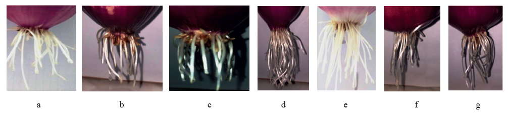
Table 2 shows a highly significant decrease in the growth of the A. cepa roots treated with sodium azide at two concentrations (50 and 100 mg/mL) at three time intervals. The data obtained from the sample treated with 50 mg/mL of sodium azide after 48 h was found insignificant (P < 0.001).
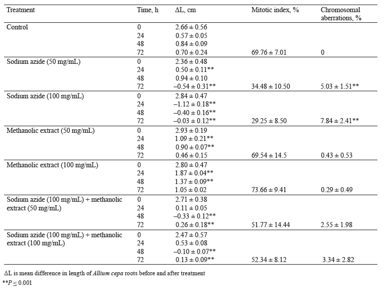
The roots treated with the methanolic extract of R. alaternus leaves showed highly significant growth (P < 0.001) after 24 and 48 h. The roots reached 8 cm after 72 h (P < 0.001) and were longer than those treated with distilled water (7 cm).
The difference in length for the antigenotoxicity test was highly significant after 48 and 72 h and not significant after 24 h. The roots demonstrated a clearly significant improvement in the diameter after 72 h. It was 0.26 and 0.13 cm, respectively, for the two extract concentrations.
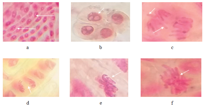
Microscopy revealed no abnormalities or disturbances in mitotic division: chromosome integrity maintained its high mitotic index (69.76 ± 7.01%), and no chromosomal aberrations were registered (Fig. 2, Table 2).

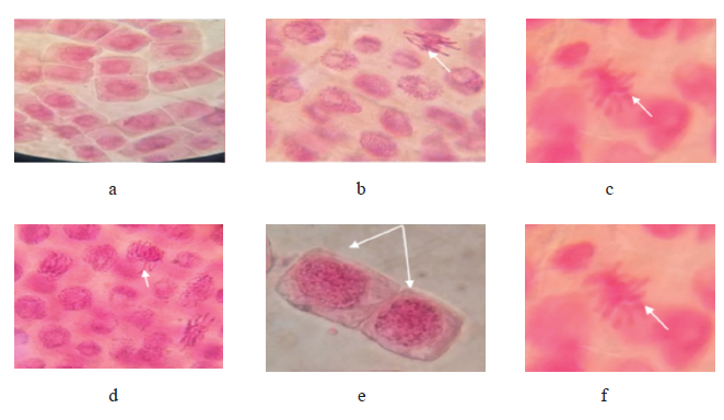
The microscopy of the roots stained with 2% acetic carmine after treatment with two concentrations of sodium azide revealed several chromosomal anomalies with disruption of all the stages of cell division (Figs. 3 and 4, Table 2). Several cells contained C-mitosis, S-mitosis, chromosomal breaks, bridges, and uneven distribution of chromosomes, which led to disturbed anaphases, metaphases, and telophases. These anomalies were caused by both concentrations of azide; however, they were much more severe at 100 mg/mL of sodium azide.
Chromosomal aberrations increased together with the concentration of sodium azide. The genotoxic effect was most severe at 100 mg/mL. Both concentrations of sodium azide reduced the mitotic index, which meant that sodium azide blocked cell division. On the other hand, the number of chromosomal aberrations grew together with sodium azide concentration. They were represented mainly by C-mitosis, chromosomal bridges and breaks, and nuclear lesions of binucleate types. Therefore, sodium azide was an aberration inducer (Figs. 3 and 4, Table 2).

Sodium azide produced a cytotoxic effect which led to poor growth and length narrowing. Its mitodepressive effect decreased mitotic activity and increased chromosomic abnormality incidence. Indeed, chemical agents are recognized as factors involved in the structural and numerical modifications of chromosomes. As a result, they cause defects in chromosome segregation, abnormal DNA replication, and DNA breaks. These chromosomal aberrations result from clastogenic and aneugenic effects [23]. This study confirmed the genotoxic effect of sodium azide. According to Al-Qurainy et al., sodium azide is a mutagenic metabolite that damages DNA by substituting one base pair with another [24]. Indeed, the shorter length of A. cepa roots treated with sodium azide could be explained by the mitodepressive effect caused by the apoptosis of meristem cells. Other samples demonstrated evolution of the normal length, probably, due to the resumption of mitosis.
Sodium azide induced the development of chromosome bridges in the meristem cells of A. cepa roots. According to Neelamkavil and Thoppil, the chromosomal aberrations and nuclear lesions in A. cepa root meristems treated with bleaching powder indicated a genotoxic effect, which confirms that sodium azide is genotoxic [25]. The clastogenic effects suggest that bleaching powder caused chromosome and chromatin breaks, which, in return, led to abnormal chromosome number, stickiness, breakage, and reunion of chromosome, as well as to bridges during mitotic division [26, 27].
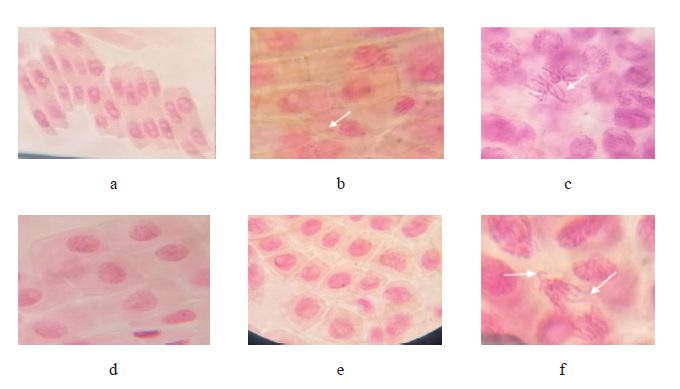
The mitotic index was higher in the roots treated with two concentrations of the R. alaternus extract than in those treated with distilled water. Therefore, the extract induced cell division and, subsequently, produced a genoprotective effect. Moreover, the number of cells in division was high with traces chromosomal aberrations also proven by a marked root length. The samples treated with 50 mg/mL of R. alaternus methanolic extract had pycnotic nuclei and chromosomal breaks (Figs. 5 and 6, Table 2). This finding confirms the conclusion made by Ben Ammar et al., who experimented with methanolic, petroleum ether, chloroform, and aqueous extracts of R. alaternus leaves and registered no mutagenicity, which means that R. alaternus is a promising antimutagenic [28].


The antigentoxic effect showed that the mitotic index was close to that of the control. It had a moderate chromosomal aberration percentage, chromosomal bridges and breaks, and a lower C-mitosis (Figs. 7 and 8, Table 2).
A quantitative analysis of the R. alaternus methanolic extract revealed a lot of polyphenols and flavonoids and thus a prominent antioxidant effect. This antioxidant effect might be the cause of the continuous cell division, mitoprotective activity, and a good DNA protection. Perron et al. tested 12 polyphenolic compounds, which demonstrated a 100% ability to inhibit DNA damage [29]. The polyphenolic compounds had hydroxyl radicals in their chemical structures, which prevented oxidative DNA damage.
On the other hand, Silva et al. showed that flavonoids have special DNA repair mechanisms that enable them to reduce and repair DNA strand breaks induced by oxidative stress [30]. Therefore, polyphenols are effective protectors against oxidative DNA damage.
Polyphenols have a powerful potency to donate electrons or hydrogen atoms, thus hampering oxidative stress, cell damage, and inflammation. They create a defensive obstacle against free radicals and reactive oxygen species. These protective effects might be explained by their antioxidant capacity [31].
Probably, the high content of flavonoids and polyphenols in R. alaternus protected the DNA as they opposed to the attack of free radicals emitted by sodium azide. Previous studies attributed the antigenotoxic activity of plant extracts to their numerous phenolic and flavonoid compounds, as well as to their ability to combat oxidative stress. The results imply that the extract managed to protect the genome of the roots against mutagen because division anomalies were very scarce. These findings confirm those made by Ben Amar et al., who proved that the extract of R. alaternus leaves and roots had antifree- radical, anti-mutagenic, and antiproliferative properties as they trapped mutagenic free radicals [32]. According to Ben Sghaier et al., medicinal plants contain phytochemicals that may have potential chemopreventive activity since they protect DNA from attack of free radicals [33].
ВЫВОДЫ
Medicinal plants contain a lot of secondary metabolites with beneficial therapeutic and pharmacological properties, which deserve extensive research. Rhamnus alaternus L. proved to be an effective antioxidant and mitoprotector that can boost the development of pharmacognosy and produce new herbal drugs for the pharmaceutical industry. The genoportective effect of R. alaternus leaf extract could be a source of new cancer drugs and protect human genome from the side effects of chemical treatment.Вклад авторов
L. Gadouche conceived and designed the analysis, performed the biological experiments, and wrote the paper. A. Zidane and K. Zerrouki contributed to the data analysis and revised the paper. A. Ababou performed the statistical analysis. I. Bachir Elazaar, D. Merabet, W. Henniche, and S. Ikhlel performed the biological experiments. All the authors revised the manuscript for publication.КОНФЛИКТ ИНТЕРЕСОВ
The authors declare that there was no potential conflict of interests regarding the publication of this article.СПИСОК ЛИТЕРАТУРЫ
- Sabahi Z, Soltani F, Moein M. Insight into DNA protection ability of medicinal herbs and potential mechanisms in hydrogen peroxide damages model. Asian Pacific Journal of Tropical Biomedicine. 2018;8(2):120–129. https://doi.org/10.4103/2221-1691.225616
- Kitagishi Y, Kobayashi M, Matsuda S. Protection against cancer with medicinal herbs via activation of tumor suppressor. Journal of Oncology. 2012;2012. https://doi.org/10.1155/2012/236530
- Dayani MA, Heidari-Soureshjani S, Salcher J. Radio-protective effects and mechanisms of medicinal plants against X-ray: A systematic review. Journal of Pharmaceutical Research International. 2019;25(1):1–11. https://doi.org/10.9734/JPRI/2018/46764
- Nekkaa A, Benaissa A, Mutelet F, Canabady-Rochelle L. Rhamnus alaternus plant: Extraction of bioactive fractions and evaluation of their pharmacological and phytochemical properties. Antioxidants. 2021;10(2). https://doi.org/10.3390/antiox10020300
- Zeouk I, Bekhti K. A critical overview of the traditional, phytochemical and pharmacological aspects of Rhamnus alaternus: a Mediterranean shrub. Advances in Traditional Medicine. 2020;20:1–11. https://doi.org/10.1007/s13596-019-00388-8
- Boussahel S, Speciale A, Dahamna S, Amar Y, Bonaccorsi I, Cacciola F, et al. Flavonoid profile, antioxidant and cytotoxic activity of different extracts from Algerian Rhamnus alaternus L. bark. Pharmacogny Magazine. 2015;11(42):S102–S109. https://doi.org/10.4103/0973-1296.157707
- Moussi K, Nayak B, Perkins LB, Dahmoune F, Madani K, Chibane M. HPLC-DAD profile of phenolic compounds and antioxidant activity of leaves extract of Rhamnus alaternus L. Industrial Crops and Products. 2015;74:858–866. https://doi.org/10.1016/j.indcrop.2015.06.015
- Ben Ammar R, Kilani S, Bouhlel I, Skandrani I, Naffeti A, Boubaker J, et al. Antibacterial and cytotoxic activities of extracts from (Tunisian) Rhamnus alaternus (Rhamnaceae). Annals of Microbiology 2007;57(3):453–460. https://doi.org/10.1007/BF03175089
- Bhouri W, Sghaier MB, Kilani S, Bouhlel I, Dijoux-Franca M-G, Ghedira K, et al. Evaluation of antioxidant and antigenotoxic activity of two flavonoids from Rhamnus alaternus L. (Rhamnaceae): Kaempferol 3-O-β-isorhamninoside and rhamnocitrin 3-O-β-isorhamninoside. Food and Chemical Toxicology 2011;49(5):1167–1173. https://doi.org/10.1016/j.fct.2011.02.011
- Bhouri W, Bouhlel I, Boubaker J, Kilani S, Ghedira K, Ghedira LC. Induction of apoptosis in human lymphoblastoid cells by kaempferol 3-O-β-isorhamninoside and rhamnocitrin 3-O-β-isorhamninoside from Rhamnus alaternus L. (Rhamnaceae). Cell Proliferation 2011;44(3):283–290. https://doi.org/10.1111/j.1365-2184.2011.00749.x
- Chatti IB, Ben Toumia I, Krichen Y, Maatouk M, Ghedira LC, Krifa M. Assessment of Rhamnus alaternus leaves extract: Phytochemical characterization and antimelanoma activity. Journal of Medicinal Food. 2021. https://doi.org/10.1089/jmf.2020.0170
- Gadouche L, Zidane A, Zerrouki K, Azouni K, Bouinoune S. Cytotoxic effect of Myrtus communis, Aristolochia longa, and Calycotome spinosa on human erythrocyte cells. Foods and Raw Materials. 2021;9(2):379–386. https://doi.org/10.21603/2308-4057-2021-2-379-386
- Quezel P, Santa S. New flora of Algeria and southern desert regions. 2 Vol. Paris: National Centre for Scientific Research; 1963. 1170 p. (In French.).
- Diallo D, Sanogo R, Yasambou H, Traoré A, Coulibaly K, Maiga A. Study of the chemical compounds of Ziziphus mauritiana Lam. (Rhamnaceace) leaves, used traditionally in the treatment of diabetes in Mali. Comptes Rendus Chimie. 2004;7(10–11):1073–1080. (In French.). https://doi.org/10.1016/j.crci.2003.12.035
- Raafat K, Samy W. Amelioration of diabetes and painful diabetic neuropathy by Punica granatum L. extract and its spray dried biopolymeric dispersions. Evidence-Based Complementary and Alternative Medicine. 2014;2014. https://doi.org/10.1155/2014/180495
- Hmid I, Elothmani D, Hanine H, Oukabli A, Mehinagic E. Comparative study of phenolic compounds and their antioxidant attributes of eighteen pomegranate (Punica granatum L.) cultivars grown in Morocco. Arabian Journal of Chemistry. 2013;10:S2675–S2684. https://doi.org/10.1016/j.arabjc.2013.10.011
- Burits M, Bucar F. Antioxidant activity of Nigella sativa essential oil. Phytotherapy Research. 2000;14(5):323–328. https://doi.org/10.1002/1099-1573(200008)14:5<323::AID-PTR621>3.0.CO;2-Q
- Sharififar F, Mozaffarian V, Moradkhani S. Comparison of antioxidant and free radical scavenging activities of the essential oils from flowers and fruits of Otostegia persica Boiss. Pakistan Journal of Biological Sciences. 2007;10(21):3895–3899. https://doi.org/10.3923/pjbs.2007.3895.3899
- Kartal N, Sokmen M, Tepe B, Daferera D, Polissiou M, Sokmen A. Investigation of the antioxidant properties of Ferula orientalis L. using a suitable extraction procedure. Food Chemistry. 2007;100(2):584–589. https://doi.org/10.1016/j.foodchem.2005.09.084
- Tedesco SB, Laughinghouse IV HD. Bioindicator of genotoxicity: The Allium cepa test. In: Srivastava JK, editor. Environmental contamination. Rijeka: InTech Publisher; 2012. pp. 137–156. https://doi.org/10.5772/31371
- Yekeen TA, Azeez MA, Akinboro A, Lateef A, Asafa TB, Oladipo IC, et al. Safety evaluation of green synthesized Cola nitida pod, seed and seed shell extracts-mediated silver nanoparticles (AgNPs) using an Allium cepa assay. Journal of Taibah University for Science. 2017;11(6):895–909. https://doi.org/10.1016/j.jtusci.2017.06.005
- Grant WF. The present status of higher plant assays for the detection of environmental mutagens. Mutation Research Regular Papers. 1994;310(2):175–185. https://doi.org/10.1016/0027-5107(94)90112-0
- Leme DM, Marin-Morales MA. Allium cepa test in environmental monitoring: A review on its application. Mutation Research – Reviews in Mutation Research. 2009;682(1):71–81. https://doi.org/10.1016/j.mrrev.2009.06.002
- Al-Qurainy F, Al-Hemaid FM, Khan S, Ajmal Ali M, Tarroum M, Ashraf M. Detection of sodium azide-induced mutagenicity in the regenerated shoots of Artemisia annua using internal transcribed spacer (ITS) sequences of nrDNA. Pakistan Journal of Botany. 2011;43(4):2183–2186.
- Neelamkavil SV, Thoppil JE. Genotoxic assessment of calcium hypochlorite and Strychnos potatorum Linn. seeds – Two commonly used water purifying agents. Reviews on Environmental Health. 2015;30(1):19–23. https://doi.org/10.1515/reveh-2014-0066
- Bhagyanathan NK, Thoppil JE. Genotoxic potential of Cynanchum sarcomedium Meve & Liede coupled with its modulatory action on oxidative-stress–mediated genotoxicity by hydrogen peroxide. Turkish Journal of Biology. 2016;40(1):120–129.
- Obute GC, Ekeke C, Izuka DC. Genotoxicity assessment of refined petroleum products and popular local soft drink (Zobo) in daily use in Nigeria. Research Journal of Mutagenesis. 2016;6(1):22–30. https://doi.org/10.3923/rjmutag.2016.22.30
- Ben Ammar R, Kilani S, Abdelwahed A, Hayder N, Mahmoud A, Chibani J, et al. In vitro mutagenicity, antimutagenicity and free radical scavenging activity of Rhamnus alaternus (Rhamnaceae) extracts. Pakistan Journal of Biological Sciences. 2005;8(3):439–445. https://doi.org/10.3923/pjbs.2005.439.445
- Perron NR, Hodges JN, Jenkins M, Brumaghim JL. Predicting how polyphenol antioxidants prevent DNA damage by binding to iron. Inorganic Chemistry. 2008;47(14):6153–6161. https://doi.org/10.1021/ic7022727
- Silva JP, Gomes AC, Coutinho OP. Oxidative DNA damage protection and repair by polyphenolic compounds in PC12 cells. European Journal of Pharmacology. 2008;601(1–3):50–60. https://doi.org/10.1016/j.ejphar.2008.10.046
- Majidinia M, Bishayee A, Yousefi B. Polyphenols: Major regulators of key components of DNA damage response in cancer. DNA Repair. 2019;82. https://doi.org/10.1016/j.dnarep.2019.102679
- Ben Ammar R, Kilani S, Bouhlel I, Ezzi L, Skandrani I, Boubaker J, et al. Antiproliferative, antioxidant, and antimutagenic activities of flavonoid-enriched extracts from (Tunisian) Rhamnus alaternus L.: Combination with the phytochemical composition. Drug and Chemical Toxicology. 2008;31(1):61–80. https://doi.org/10.1080/01480540701688725
- Ben Sghaier M, Bhouri W, Neffati A, Boubaker J, Skandrani I, Bouhlel S, et al. Chemical investigation of different crude extracts from Teucrium ramosissimum leaves. Correlation with their antigenotoxic and antioxidant properties. Food and Chemical Toxicology. 2011;49(1):191–201. https://doi.org/10.1016/j.fct.2010.10.016
