

Аннотация
The olive (Olea europaea L.) is one of the most important plants grown in many Mediterranean countries that has a high economic value. Olives, which are specific to each region, have different bioactive components. In this study, we investigated the phenolic/flavonoid contents, as well as antioxidant, antimicrobial, and antithrombotic activities of the fruit, leaf, and seed extracts obtained from the Halhalı olive grown in Arsuz district of Hatay, Turkey.Antioxidant activities of the phenolic compounds found in the olive fruit, seed, and leaf extracts were determined by employing established in vitro systems. Total phenolics were determined as gallic acid equivalents, while total flavonoids were determined as quercetin equivalents. Also, we evaluated a possible interaction between oleuropein and aggregation-related glycoproteins of the platelet surface via docking studies.
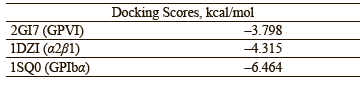
The extracts showed effective antioxidant activity. The seed extract had the highest phenolic content of 317.24 μg GAE, while the fruit extract had the highest flavonoid content of 4.43 μg. The highest potential for metal chelating activity was found in the leaf extract, with an IC50 value of 13.33 mg/mL. Also, the leaf extract showed higher levels of antioxidant, antithrombotic, and antimicrobial activity, compared to the fruit and seed extracts. The docking scores of oleuropein against the target molecules GPVI, α2β1, and GPIbα were calculated as –3.798, –4.315, and –6.464 kcal/mol, respectively.
The olive fruit, leaf, and seed extracts used as experimental material in our study have remarkable antioxidant, antimicrobial, and antithrombotic potential.
Ключевые слова
Olea europaea L., antioxidant, antithrombotic activity, antimicrobial activity, molecular dockingВВЕДЕНИЕ
The olive (Olea europeae L.), the oldest fruit tree known to be cultivated in the world, belongs to the Olea genus of the Oleaceae family and takes its name from the Greek “elaia” and the Latin “olea”. People consume its fruit and other products for both nutritional and health benefits [1–3].
Like most plants, the olive tree is constantly exposed to environmental stresses such as high temperatures and UV radiation. To protect itself in this situation, the plant produces chemical components with antioxidant properties defined as phenolic compounds (phenolic acids, phenolic alcohols, secoiridoids, and flavonoids). These components vary qualitatively and quantitatively depending on certain factors such as the olive species, the degree of its fruit/leaf’s ripening, the climate, and the geographical location [4–8]. Phenolic compounds are found in the olive’s fruit, leaves, and seeds. They contribute to the color and flavor of the fruit depending on their concentration, which differs considerably between various tissues [9, 10].
Olive groves, which cover 8 million hectares of Mediterranean countries, have a very important social and economic value. Olive oil is the main product produced by these countries on a large scale. However, the by-products of such production are used beneficially, too. They include olive cake (olive seed and pomace, the fleshy part), black water, twigs, and leaves.
There is a lot of research on olive leaves and seeds, which have been used in folk medicine for a long time but have become more popular recently. Olive leaves are commercially produced in the form of herbal tea and olive leaf extract is offered in the form of tablets, both considered as food support products. In cosmetics, products containing olive leaf extract are used in skin care due to their antioxidant and antiaging effects.
Recently, some medicines made from olive leaf extract have started to be used for human and animal health [11, 12]. These medicines have natural antibiotic and antiparasitic effects [13]. Olive leaves contain polyphenols (oleuropein, hydroxytyrosol, and luteolin) that can inhibit platelet aggregation and eicosanoid production [14]. Olive leaf extract has an antimicrobial effect against all kinds of bacteria, viruses, yeasts, and fungi. It is used in the treatment of common bacterial infections such as bronchitis and tonsillitis, fungal infections in the vaginal areas of women, and viral infections such as herpes [15–19].
The olive seed, which is the main waste of olive oil and table olive production, is a lignocellulosic structure made up of hemicellulose, cellulose, and lignin. It contains oil that is rich in polyunsaturated fatty acids. The amount of protein in the seed is higher than in the remaining part of the olive [20]. In addition, it contains different polyphenols such as tyrosol, hydroxytyrosol, oleuropein, and 3,4-DHFEA-EDA. The olive seed is also used for therapeutic purposes in folk medicine practices. In particular, to treat rheumatic pain or accelerate wound healing, the olive is crushed with its seed and applied onto the lesion. It can also be used for gastritis and stomach ulcers.
According to recent research, oleuropein is the most active polyphenolic antioxidant in the olive’s fruit, seed, and leaves [21, 22]. This compound was discovered as early as 1908 by Bourquelot and Vintilesco in a study on the olive’s fruit, but its structure was only identified in 1960 [23]. Most researchers agree that the therapeutic properties of olives and their by-products come from oleuropein, the most abundant phenolic compound in their composition [24–26].
The value of the olive tree lies not only in its fruit, but also in its oil, seeds, branches, roots, and leaves. Therefore, in this study, we aimed to determine phenolic components and antioxidant, antimicrobial, and antithrombotic activities of the fruit, leaf, and seed extract of the olive tree (O. europaea L.).
In addition, we evaluated a possible interaction between oleuropein and aggregation-related glycoproteins of the platelet surface via a docking study. The fruit, leaves, and seeds were collected from the Halhalı olive tree growing in the Hatay region (Turkey) during the harvest period.
ОБЪЕКТЫ И МЕТОДЫ ИССЛЕДОВАНИЯ
Plant materials. For the experiments, olives and olive leaves were collected during the harvest period from the Halhalı olive trees growing in Arsuz District of Hatay, Turkey.
Preparation of samples. Prior to analysis, the leaf samples were cleared of impurities with distilled water and dried in the shade on blotting paper. When the leaves became brittle, they were pounded in a mortar and ground into powder. The seeds were mechanically separated from the fleshy part of the fruit, washed with distilled water, laid on drying paper, and dried overnight. The dried seeds were crushed by pounding in a mortar and turned into a slurry owing to a very small amount of gel-like substances. The olives were separated from their seeds, pounded in a mortar, and then turned into a slurry using an IKA T25 knife homogenizer. On completion of the preliminary preparations, the samples were made ready for Soxhlet extraction.
Soxhlet extraction. In order to obtain extracts from the prepared samples, 30 g of raw olive fruit, 16 g of dried olive leaves, and 40 g of dried olive seeds were placed in the cartridge of the Soxhlet apparatus. Each sample was first extracted at 55°C for 3 h using 400 mL of petroleum ether, with non-polar lipophilic components removed. Afterwards, the extraction process was continued with 400 mL of ethanol used as a solvent. For this, ethanol was evaporated under vacuum in a rotary evaporator, and the high-density ethanol extracts remaining in the balloon were weighed. The prepared extracts were dissolved in distilled water to obtain stock solutions for the measurements. These stock solutions were divided into 10 equal volumes, each poured into tubes with plastic caps and stored in a deep freezer at –40°C until the measurements.
Methods for quantitative determination. In this study, all the measurements for each parameter were performed in duplicate and the data were presented as their arithmetic means.
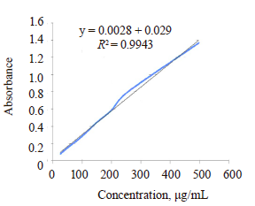
Determination of total phenolic content. The total amount of phenolic substances contained in the three extracts (fruit, seed, and leaf) was determined according to the method of Slinkard and Singleton using the Folin-Ciocaltaeu reagent [27]. Gallic acid was used as a standard. Total phenols were calculated as gallic acid equivalents using a standard calibration curve obtained from the absorbance values read against the gallic acid solutions in ethanol at 25, 50, 100, 150, 200, 250, and 500 μg/mL concentrations. Spectrophotometric and absorbance measurements were performed at 765 nm on a Shimadzu 1201 device.
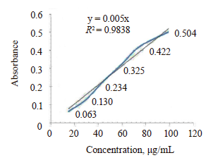
Determination of total flavonoid content. The total flavonoid content in the samples was determined according to the method of Moreno et al., which was based on the formation of complexes of flavonoid groups with metal ions [28]. In this study, we used aluminum as a metal ion and quercetin as a standard molecule. A stock solution was prepared with a final concentration of quercetin in methanol reaching 100 μg/mL. Dilute solutions with concentrations of 15, 30, 45, 60, and 75 μg/mL were prepared from this solution. For this, we placed 100 μL of 10% AI(NO3)3 in the test tubes, first adding 100 μL of 1 M CH3COOK and 3.8 mL of methanol, and then adding 1 mL of the standard/extract solutions. The mixture was vortexed and incubated for 45 min in a shaking water bath at 25°C. The absorbance of the formed yellow complex was measured at 415 nm in the spectrophotometer. Total flavonoids were calculated as quercetin equivalents with the help of the quercetin standard curve.
Determination of reducing capacity. The Oyaizu method was used to determine the reducing capacity of the samples [29]. This method is based on monitoring the absorbance at 700 nm for the yellow color that turns green in the experimental environment with the Fe3+/Fe2+ conversion, with high absorbance corresponding to high reduction potential. The solutions with 5, 10, 25, 50, 100, and 150 μg/mL concentrations of butylated hydroxyanisole were used as standards. The standard graph was drawn using the absorbances of the butylated hydroxyanisole standards spectrophotometrically measured at 700 nm. The reducing potential of the samples was determined using this calibration graph.
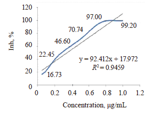
Determination of metal ion chelating activity. The chelating of ferrous ions by the ethanol extracts of Halhali olives was estimated by the method of Dinis et al. [30]. For this, the extracts were added to a solution of 2 mM FeCl2. The reaction was initiated by adding 5 mM of ferrozine, and the mixture was shaken vigorously and left standing at room temperature for 10 min. The absorbance of the solution was then measured spectrophotometrically at 562 nm. Ethylenediamine tetraacetic acid (EDTA) was used as a positive control. The percentage of inhibition of the ferrozineFe2+ complex formation was calculated using the formula below:
![]()
where A0 is the absorbance of the control; A1 is the absorbance in the presence of the extract samples or standards.
Determination of H2O2 scavenging activity. The hydrogen peroxide scavenging activity was determined according to the titration method developed by Zhang [31]. Ascorbic acid was used as a positive control. The sample or the positive control (at concentrations ranging from 50 to 1000 µg/mL) and H2O2 (0.10 mM) were added to an Erlenmeyer flask for the titration reaction. Ammonium molybdate (3%), H2SO4 (2.0 M), and KI (1.8 M) were added and mixed. The resulting yellow mixture was titrated with 5 mM Na2S2O3 until the yellow color disappeared. The hydrogen peroxide removal activity (%) of the extracts and standard substances was calculated according to the formula below:
![]()
where V0 is the control titrant volume; V1 is the sample and standard titrant volume.
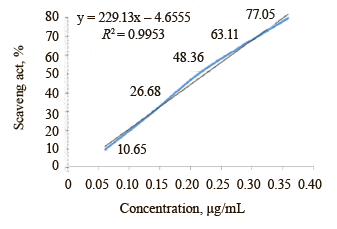
Determination of DPPH radical quenching activity. The 2,2-diphenyl-1-picrylhydrazyl (DPPH) radical quenching activity was determined according to the method of Blois [32]. This long-established method evaluates the potential of DPPH, which is a stable free radical, to react with phenolic molecules with hydrogen donor properties in the reaction medium. The DPPH concentration decreased due to the reaction can be monitored spectrophotometrically at 517 nm. Butylated hydroxyanisole was used as a positive control. Dilute solutions of the standard at concentrations of 5, 10, 25, 50, 100, and 150 μg/mL were prepared. The 0.1 mM solution of DPPH was prepared in 70% methanol. 3 mL of the standard/sample solution was taken and placed in test tubes. After adding 1 mL of the DPPH solution, the mixture was thoroughly vortexed and incubated for 30 min at room temperature in the dark. At the end of the incubation, absorbance was measured in the spectrophotometer at 517 nm. The radical scavenging effects (%) of the solutions were calculated using the equation below based on the absorbance values compared to the DPPH solution used as a control:
![]()
The solution prepared by mixing only 3 mL of methanol and 1 mL of DPPH solution without adding any chemicals was used as a control solution. A0 is the absorbance of control; A1 is the absorbance of sample/standard.
Determination of antimicrobial activity. The antimicrobial activity of the olive leaf, fruit, and seed extracts was determined using the agar dilution method recommended by the Clinical and Laboratory Standards Institute. Minimal inhibitory concentrations (MICs) for each extract were tested against standard bacterial (Staphylococcus aureus ATCC 29213, Enterococcus faecalis ATCC 29212, S. aureus MRSA ATCC 43300, Escherichia coli ATCC 25922, Klebsiella pneumoniae ATCC 700603, Pseudomonas aeruginosa ATCC 27853, and Acinetobacter baumannii ATCC 19606) and fungal (Candida glabrata ATCC 90030 and Candida albicans ATCC 14053) strains. The strains were obtained from the American Type Culture Collection (Rockville, USA). The bacterial strains were grown in a Mueller Hinton Broth (Merck), while the fungal strains were grown in a RPMI 1640 Broth (Sigma-Aldrich Chemie GmbH Taufkirchen, Germany).
To obtain a standard inoculum, the turbidity of bacteria and fungi was prepared according to the Mcfarland 0.5 chart. All dilutions were made with distilled water. The final concentrations of the extracts were diluted to 800, 400, 200, 100, 50, 25, 12.5, and 6.25 μg/mL. Fluconazole was used as a standard drug for fungi, while ampicillin, ciprofloxacin, amikacin, ampicillin, tigecyline, and vancomycin were used as standard drugs for bacteria. Standard inoculums of bacteria and fungi (106 CFUs/mL) were inoculated onto agar plates with a sterile plastic ring-tipped loop (0.01 mL). The plates were evaluated after they were kept in an oven at 35°C for 16–20 h for bacteria and 48 h for fungi. Minimal inhibitory concentrations were determined as the lowest concentrations that inhibited the growth of bacteria and fungi [33, 34].
Determination of antithrombotic activity. The antithrombotic activity of the extracts was determined by using a Chronolog aggregometer and the optical aggregometry technique [35]. Optical aggregometers are modified spectrophotometric instruments. They measure changes in light transmittance through the cuvette by adding a stimulating agent (an agonist such as collagen or ADP) while the platelet-rich plasma is mixed at a certain speed and the platelets form aggregates. The system calibrates itself to detect 100 units of transmission difference between the platelet-poor plasma and the platelet-rich plasma.
In our experiments, 75 mL of venous blood taken from a 47-year-old healthy male after 8 h of fasting was poured into tubes with EDTA, 3 mL for each tube. The platelet-poor plasma was obtained by centrifugation at 1000 rpm for 10 min, and the platelet-rich plasma was obtained by centrifuging at 3500 rpm for 10 min. For all the measurements, 950 µL of these plasmas was added to the measuring cuvettes. For the blank measurement, only the platelet-rich plasma was added (without any extract), and the aggregation was measured after adding the agonist (collagen). For the sample measurements, the extracts were added to the platelet-rich plasma samples at varying concentrations and incubated at 37°C for 5 min. The aggregation measurement was performed by adding the agonist at the end of the incubation. All the measurements were carried out in duplicate to calculate the averages. On completion of the measurements, the percentage of aggregation inhibition corresponding to a concentration of 1 mg/mL was calculated for each extract according to the formula below:
![]()
where Tk is the transmission of control, %; T1 is the transmission of sample, %.
Docking studies. The Maestro 12.8 (Schrödinger, New York) program was used in all molecular docking studies. The ligand structures were prepared with the 2D Sketcher. The ligands were minimized using the LigPrep, a utility of the Schrödinger software. The crystal structure of human platelet Glycoprotein VI (GPVI, pdb id: 2GI7), integrin alpha2 I domain/collagen complex (α2β1, pdb id: 1DZI), and the complex of the wild-type von Willebrand factor A1 domain and Glycoprotein Ib alpha (GPIbα, pdb id: 1SQ0) were downloaded from the RCSB Protein Data Bank (www. rcsb.org) [36–38].
Schrödinger’s modules (Protein Preparation Wizard Prime, Impact, Epik, Propka, and Prime) were used to remove the ligands and solvent molecules in protein, add hydrogens, assign charges, and delete polar hydrogens for clarity. After the target region of the proteins was determined, a grid box was created with the grid generation panel. Then, the prepared ligands were docked in this grid map 50 times in the standard precision mode using the Glide software [39].
РЕЗУЛЬТАТЫ И ИХ ОБСУЖДЕНИЕ
Oxidative damage is involved in the etiopathogenesis of many diseases and antioxidants play an important role in preventing the formation and progression of many diseases [40]. Plants, especially those used as natural resources, provide a diverse set of broad substrates for drug discovery. They have therapeutic properties due to the presence of phytochemicals with antioxidant properties such as phenols, flavonoids, and sterols.
In our study, we aimed to investigate the pharmaceutical potential of the Halhali olive’s leaf, fruit, and seed extracts using several different methods. For this purpose, we compared the antioxidant, antimicrobial, and antithrombotic activities of the extracts, and tested the interaction of oleuropein, the most effective molecule in olive composition, with the platelet surface receptors GPVI, α2β1, and GPIbα by the docking technique.
The phenolic contents of the extracts were calculated as gallic acid equivalents (µg gallic acid/mg extract) using the standard graph (Fig. 1). Similarly, the flavonoid contents were calculated as quercetin equivalents (µg quercetin/mg extract) using the standard graph (Fig. 2). The total phenolic and flavonoid equivalent substance contents of the extracts are given in Table 1.

The total phenolic content of the fruit extract was found to be lower than that of the leaf and seed extracts, while its total flavonoid content was the highest at 4.43 μg, compared to 4.24 and 0.86 μg in the leaf and seed extracts, respectively. The percentage of flavonoids in the total phenolic content was determined as 1.51% for the leaf extract, 1.63% for the fruit extract, and 0.27% for the seed extract. These values show that the jelly-oily component, which is found in the interior of the olive pit and contains bioactive components in the olive pit extract, is rich in phenolics. However, its percentage of flavonoids in total phenolics is lower compared to that in the leaf and fruit extracts. Çetinkaya and Kulak, in their study of olives from the Kilis region, reported that the ratios between the total phenolic content and the flavonoid composition of this content differed depending on the development period of the olive [41].
The free radical scavenging effect of all the extracts was measured by DPPH assays. The DPPH radical quenching activity was determined according to the method of Blois [32]. The calibration curve was prepared using diluted solutions of 5, 10, 25, 50, 100, and 150 μg/mL of butylated hydroxyanisole, a strong radical scavenging antioxidant used as a standard.
The IC50 values calculated for the DPPH radical scavenging activity were 9.22 μg/mL for butylated hydroxyanisole and 3.80, 9.47, and 5.25 mg/mL for the leaf, fruit, and seed extracts, respectively. The antiradical activity values defined as 1/IC50 were 0.26, 0.11, and 0.19 for the leaf, fruit, and seed extracts, respectively. The strongest DPPH radical scavenging effect was observed in the leaf extracts, followed by the seed and fruit extracts.
Martinez et al. reported that olive fruit extracts had strong radical scavenging activity against the DPPH radical [42]. Stankovich et al., who used the DPPH method to determine antioxidant activity in olive leaf extracts from France and Serbia, found IC50 values of 113.30 and 94.39 μg/mL, respectively [43]. Orak et al. reported the IC50 values in Çekişte and Uslu olive leaf extracts to reach 0.63 and 0.65 mg/mL, respectively [44].
The reducing potential of Fe(III) to Fe(II) of the olive leaf, fruit, and seed extracts was determined according to the Oyaizu method [29]. Butylated hydroxyanisole was used as a standard molecule. According to our findings, the reducing potentials of 1 mg/mL extracts in terms of butylated hydroxyanisole equivalents were 257.6, 77.25, and 21.24 μg/mL for the leaves, fruit, and seeds, respectively. We observed the strongest reducing potential in the seed extract. Altemimi et al., who tested antioxidant parameters in methanol and ethanol extracts of olive leaves, found that methanol extracts had the highest phenolic content, while ethanol extracts had higher reducing potential [45]. Fu et al. reported the ferric reduction value of the extractable components of olive samples to be 2.70 ± 0.03 μmol Fe(II)/g [46].
The metal chelating potentials of the olive fruit, leaf, and seed extracts and the standard chelator (EDTA) were determined according to Dinis method [30]. The % inhibition-concentration graph (Fig. 3) was created by using dilute solutions of EDTA at varying concentrations.
According to our findings, the highest metal chelating activity (IC50) was seen in the leaf extract (13.33 mg/mL), followed by the seed and fruit extracts (17.37 and 24.76 mg/mL, respectively). Wang et al. reported that neuron damage due to lead toxicity in the brain was significantly prevented, and antioxidant capacity increased, in the mice that received olive leaf extracts [47]. Fabiani et al. suggested that olive phenolic components protected human mononuclear blood cells and reduced oxidative damage due to their metal chelating abilities among other factors [48].
The hydrogen peroxide scavenging activity was determined according to the method of Zhang et al. [31]. Ascorbic acid was used as a standard molecule. We observed that the leaf extract had the strongest activity, with an IC50 of 4.27 mg/mL, while the values for the fruit and seed extracts were 36.39 and 30.50 mg/mL, respectively (Fig. 4). Lins et al. reported that olive leaf extracts showed protective activity against oxidative stress on erythrocytes in vitro, inhibiting the lysis of erythrocytes and reducing MDA levels formed by oxidation of erythrocyte membrane lipids [49].
The antimicrobial activity of the olive leaf, fruit, and seed extracts was determined using the agar dilution method recommended by the Clinical and Laboratory Standards Institute [33, 34]. The lowest effective concentrations that prevented the growth of bacteria and fungi were determined as minimal inhibitory concentrations (MICs). They were tested against the standard bacterial and fungal strains. Fluconazole was used as a standard drug for fungi, while ampicillin, ciprofloxacin, amikacin, ampicillin, tigecyline, and vancomycin were used as standard drugs for bacteria. The extracts showed varying degrees of antimicrobial and antifungal activity against all tested microorganisms. However, the leaf extract showed stronger antimicrobial and antifungal effects (100–200 μg/mL) compared to the fruit and seed extracts (200–400 μg/mL).
While the leaf extracts were similarly effective on all the tested microorganisms, the fruit and seed extracts were found to be less effective against Pseudomonas aeruginosa, Acinetobacter baumannii, and MRSA, which are most commonly isolated from hospital infections (400–800 μg/mL).
Pereira et al. analyzed the phenolic compounds in the aqueous extract of powdered olive leaves using HPLC/DAD and investigated their antimicrobial properties. They reported that the inhibitory effects of different concentrations of the obtained extract on microorganisms were respectively found as Bacillus cereus ~ Candida albicans > Escherichia coli > Staphylococcus aureus > Cryptococcus neoformans ~ Klebsiella pneumoniae ~ P. aeruginosa > Bacillus subtilis [50].
The antithrombotic activity was determined by using the Chronolog system and the turbidimetry technique. According to our data, all three extracts showed antithrombotic activity at certain rates, but the leaf extract had higher values compared to the fruit and seed extracts. The relative antithrombotic activities of the extracts were 23.22, 5.01, and 7.36% for the leaves, fruit, and seeds, respectively, with the control activity of 100%.
Dub and Dugani, in their study on rabbits, reported that the application of repeated amounts of olive leaf extracts reduced thrombus formation and the potential for attachment to the vessel wall by changing the thrombus morphology [51]. Gorzynik-Debicka et al. found that olives and olive oil had antiatherogenic and antithrombotic effects, as well as anticarcinogenic effects [52]. Zbidi et al. showed that oleuropein and (+)-cycloolivil molecules isolated from the olive tree had strong antithrombotic effects [53]. Similarly, Petroni et al. reported that polyphenolic compounds extracted from olives inhibited eicosanoid production and platelet aggregation [54].
The molecular docking technique evaluates the interaction of a molecule with the binding site of an enzyme or a receptor with a protein structure. Certain score algorithms simulate the placement of a molecule in the protein structure by taking into account many factors, including the electro negativities of the atoms, their positions to each other, and the conformation of the molecule to be placed into the protein structure.
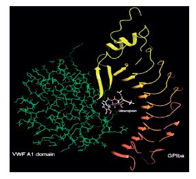
In this study, we selected platelet adhesion receptors GPVI, α2β1, and GPIbα as potential targets and investigated the docking scores and interactions of oleuropein, the olive’s major bioactive component, with these targets [55]. We calculated the docking scores of oleuropein against the target molecules (Table 2).
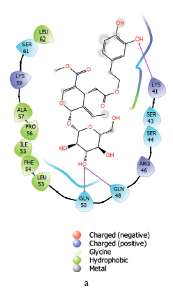
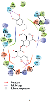
Oleuropein hydrogen bonded with LYS 41, GLN 50, and GLN 48 in the epitope part of the active site of GPVI. It interacted hydrophobicly with ALA 57, PRO 56, ILE 55, PHE 54, and LEU 53, and had polar interactions with SER 43, SER 44, and SER 61. Oleuropein established a metal coordination bond with Co2+ in the active site of the α2β1 protein. Oleuropein hydrogen bonded with GLU 256, SER 257, and GLU 299. The molecule interacted hydrophobically with LEU 220, PHE 224, and LEU 296, and polarly with SER 153, ASN 154, SER 155, THR 221, SER 257, HIE 258, and ASN 295. Oleuropein hydrogen bonded with ARG 64, ASP 83, ASP 106, LYS 132, and GLN 604 in the active site of GPIbα. It also interacted with ASN 61, SER 85, HIS 86, SER 108, SER 154, GLN 232, THR 176, and VAL 104. It interacted polarly with PHE 109, TRP 230, ILE 605, TYR 600, MET 239, LEU 178, TYR 130, and LEU 131 (Figs 5 and 6).

ВЫВОДЫ
Since oxidant/antioxidant imbalance is extremely dangerous for the organism and new side effects of synthetic antioxidants are revealed every day, the value of natural antioxidants cannot be overestimated. Our study showed that although the olive leaf extract was more effective, all the extracts (fruit, seed, and leaf) had remarkable antioxidant, antimicrobial, and antithrombotic effects. Our results confirmed their traditional use in the treatment of various disorders. This should promote the potential use of Halhalı olive leaves as a nutraceutical and pharmacological agent. Further studies can be conducted to isolate and characterize bioactive components in olive leaves, fruits, and seeds, as well as to reveal various drug candidate moleculesВклад авторов
We declare that this work was done by the authors named in this article and all liabilities pertaining to claims relating to the content of this article will be borne by the authors. F. Küçükbay designed the study and supervised the data collection. K. Batçıoğlu, M. A. Alagöz, Y. Yilmaztekin, and S. Günal analyzed and interpreted the data. All of the authors prepared the manuscript for publication and reviewed the draft. All the authors have read and approved the manuscript.КОНФЛИКТ ИНТЕРЕСОВ
The authors declare no conflict of interest associated with this workФИНАНСИРОВАНИЕ
The authors thank Inönü University (İNÜ), Turkey (BAPB – Grant No. TYL-2016-143) for financial support.СПИСОК ЛИТЕРАТУРЫ
- Parvaiz M, Hussain K, Shoaib M, William G, Tufail M, Hussain Z, et al. A review: Therapeutic significance of olive Olea europaea L. (oleaceae family). Global Journal of Pharmacy and Pharmacology. 2013;7(3):333–336.
- Ozturk M, Altay V, Gönenç TM, Unal BT, Efe R, Akçiçek E, et al. An overview of olive cultivation in Turkey: Botanical features, eco-physiology and phytochemical aspects. Agronomy. 2021;11(2). https://doi.org/10.3390/agronomy11020295
- Owen RW, Mier W, Giacosa A, Hull WE, Spiegelhalder B, Bartsch H. Phenolic compounds and squalene in olive oils: The concentration and antioxidant potential of total phenols, simple phenols, secoiridoids, lignansand squalene. Food and Chemical Toxicology. 2000;38(8):647–659. https://doi.org/10.1016/s0278-6915(00)00061-2
- Ben Mansour-Gueddes S, Saidana-Naija D, Bchir A, Braham M. Climate change effects on phytochemical compounds and antioxidant activity of Olea europaea L. Notulae Botanicae Horti Agrobotanici Cluj-Napoca. 2020;48(1):436–455. https://doi.org/10.15835/nbha48111615
- Helvacı M, Akdaş M, Özden Ö. Occurrence, damage, and population dynamics of the olive fruit fly (Bactrocera oleae Gmelin) in the Turkish Republic of Northern Cyprus. Turkish Journal of Agriculture and Forestry. 2018;42(6):453–458. https://doi.org/10.3906/tar-1802-101
- Arji I, Arzani K. Effect of water stress on some biochemical changes in leaf of five olive (Olea europaea L.) cultivars. Acta Horticulturae. 2008;791:523–526. https://doi.org/10.17660/ActaHortic.2008.791.80
- Brahmi F, Mechri B, Dabbou S, Dhibi M, Hammami M. The efficacy of phenolic compounds with different polarities as antioxidants from olive leaves depending on seasonal variations. Industrial Crops and Products. 2012;38(1):146–152. https://doi.org/10.1016/j.indcrop.2012.01.023
- Yorulmaz A, Poyrazoğlu ES, Özcan MM, Tekin A. Phenolic profiles of Turkish olives and olive oils. European Journal of Lipid Science and Technology. 2012;114(9):1083–1093. https://doi.org/10.1002/ejlt.201100186
- Soler-Rivas C, Espín J C, Wichers H J. Oleuropein and related compounds. Journal of the Science of Food and Agriculture. 2000;80(7):1013–1023. https://doi.org/10.1002/(SICI)1097-0010(20000515)80:7<1013::AID-JSFA571>3.0.CO;2-C
- Sabry OMM. Review: Beneficial health effects of olive leaves extracts. Journal of Natural Sciences Research. 2014;4(19).
- Şahin S, Bilgin M. Olive tree (Olea europaea L.) leaf as a waste by-product of table olive and olive oil industry: A review. Journal of the Science of Food and Agriculture. 2018;98(4):1271–1279. https://doi.org/10.1002/jsfa.8619
- Markhali FS, Teixeira JA, Rocha CMR. Olive tree leaves-A source of valuable active compounds. Processes. 2020;8(9). https://doi.org/10.3390/pr8091177
- Durlu-Özkaya F, Ozkaya MT. Oleuropein using as an additive for feed and products used for humans. Journal of Food Processing and Technology. 2011;2(3). https://doi.org/10.4172/2157-7110.1000113
- Singh I, Mok M, Christensen A-M, Turner AH, Hawley JA. The effects of polyphenols in olive leaves on platelet function. Nutrition, Metabolism and Cardiovascular Diseases. 2008;18(2):127–132. https://doi.org/10.1016/j.numecd.2006.09.001
- Vogel P, Machado IK, Garavaglia J, Zani VT, de Souza D, Dal Bosco SM. Polyphenols benefits of olive leaf (Olea europaea L) to human health. Nutricion Hospitalaria. 2015;31(3):1427–1433. https://doi.org/10.3305/nh.2015.31.3.8400
- Borjan D, Leitgeb M, Knez Ž, Hrnčič MK. Microbiological and antioxidant activity of phenolic compounds in olive leaf extract. Molecules. 2020;25(24). https://doi.org/10.3390/molecules25245946
- Lorzadeh N, Kazemirad Y, Kazemirad N. Treatment of genital herpes using olive leaf extract. Clinical Case Reports. 2021;9(2):986–989. https://doi.org/10.1002/ccr3.3723
- Ghanbari R, Anwar F, Alkharfy KM, Gilani A-H, Saari N. Valuable nutrients and functional bioactives in different parts of olive (Olea europaea L.) – A review. International Journal of Molecular Sciences. 2012;13(3):1291–1340. https://doi.org/10.3390/ijms13033291
- Hashmi MA, Khan A, Hanif M, Farooq U, Perveen S. Traditional uses, phytochemistry, and pharmacology of Olea europaea (olive). Evid – Based Complement and Alternative Medicine. 2015;2015. https://doi.org/10.1155/2015/541591
- Rodríguez G, Lama A, Rodríguez R, Jiménez A, Guillén R, Fernández-Bolaños J. Olive stone an attractive source of bioactive and valuable compounds. Bioresource Technology. 2008;99(13):5261–5269. https://doi.org/10.1016/j.biortech.2007.11.027
- Panizzi L, Scarpati ML, Oriente G. The constitution of oleuropein, a bitter glucoside of the olive with hypotensive action. Gazzetta Chimica Italiana. 1960;90:1449–1485.
- Kiritsakis K, Kontominas MG, Kontogiorgis C, Hadjipavlou-Litina D, Moustakas A, Kiritsakis A. Composition and antioxidant activity of olive leaf extracts from Greek olive cultivars. Journal of the American Oil Chemists' Society. 2010;87(4):369–376. https://doi.org/10.1007/s11746-009-1517-x
- Omar SH. Oleuropein in olive and its pharmacological effects. Scientia Pharmaceutica. 2010;78(2):133–154. https://doi.org/10.3797/scipharm.0912-18
- Yuan J-J, Wang C-Z, Ye J-Z, Tao R, Zhang Y-S. Enzymatic hydrolysis of oleuropein from Olea europea (olive) leaf extract and antioxidant activities. Molecules. 2015;20:2903–2921. https://doi.org/10.3390/molecules20022903
- Talhaoui N, Taamalli A, Gómez-Caravaca AM, Fernández-Gutiérrez A, Segura-Carretero A. Phenolic compounds in olive leaves: Analytical determination, biotic and abiotic influence, and health benefits. Food Research International. 2015;77:92–108. https://doi.org/10.1016/j.foodres.2015.09.011
- Antoniou C, Hull J. The anti-cancer effect of Olea europaea L. products: A review. Current Nutrition Reports. 2021;10:99–124. https://doi.org/10.1007/s13668-021-00350-8
- Slinkard K, Singleton VL. Total phenol analysis: Automation and comparison with manual methods. American Journal of Enology and Viticulture. 1977;28(1):49–55.
- Moreno MIN, Isla MI, Sampietro AR, Vattuone MA. Comparison of the free radical-scavenging activity of propolis from several regions of Argentina. Journal of Ethnopharmacology. 2000;71(1–2):109–114. https://doi.org/10.1016/s0378-8741(99)00189-0
- Oyaizu M. Studies on product of browning reaction prepared from glucose amine. Japan Journal of Nutrition. 1986;44(6):307–315. https://doi.org/10.5264/eiyogakuzashi.44.307
- Dinis TCP, Madeira VMC, Almedia LM. Action of phenolic derivatives (acetaminophen, salicylate, and 5-aminosalicylate) as inhibitors of membrane lipid peroxidation and as peroxyl radical scavengers. Archives of Biochemistry and Biophysics. 1994;315(1):161–169. https://doi.org/10.1006/abbi.1994.1485
- Zhang X-Y. Principle of chemical analysis. Beijing: China Science Press; 2000. pp. 275–276.
- Blois MS. Antioxidant determinations by the use of a stable free radical. Nature. 1958;181(4617):1199–1200. https://doi.org/10.1038/1811199a0
- M45-A2 Methods for Antimicrobial dilution and disk susceptibility testing of infrequently isolated or fastidious bacteria; Approved guideline – Second edition. Clinical Laboratory Standards Institute; 2010. 77 p.
- M100-S23 Performance standards for antimicrobial susceptibility testing; Twenty-third informational supplement. Clinical Laboratory Standards Institute; 2013. 205 p.
- Hayward CPM, Moffat KA, Raby A, Israels S, Plumhoff E, Flynn G, et al. Development of North American consensus guidelines for medical laboratories that perform and interpret platelet function testing using light transmission aggregometry. American Journal of Clinical Pathology. 2010;134(6):955–963. https://doi.org/10.1309/AJCP9V3RRVNZMKDS
- Damaskinaki F-N, Moran LA, Garcia A, Kellam B, Watson SP. Overcoming challenges in developing small molecule inhibitors for GPVI and CLEC-2. Platelets. 2021;32(6):744–752. https://doi.org/10.1080/09537104.2020.1863939
- Bivi N, Hu H, Chavali B, Chalmers MJ, Reutter CT, Durst GL, et al. Structural features underlying raloxifene’s biophysical interaction with bone matrix. Bioorganic and Medicinal Chemistry. 2016;24(4):759–767. https://doi.org/10.1016/j.bmc.2015.12.045
- Sable R, Jois S. Surfing the protein-protein interaction surface using docking methods: Application to the design of PPI inhibitors. Molecules. 2015;20(6):11569–11603. https://doi.org/10.3390/molecules200611569
- Kuzu B, Hepokur C, Alagoz MA, Burmaoglu S, Algul O. Synthesis, biological evaluation and in silico studies of some 2-substituted benzoxazole derivatives as potential anticancer agents to breast cancer. ChemistrySelect. 2022;7(1). https://doi.org/10.1002/slct.202103559
- Liang W, He X, Bi J, Hu T, Sun Y. Role of reactive oxygen species in tumors based on the “seed and soil” theory: A complex interaction (Review). Oncology Reports. 2021;46(3). https://doi.org/10.3892/or.2021.8159
- Çetinkaya H, Kulak M. Relationship between total phenolic, total flavonoid and oleuropein in different aged olive (Olea europaea L.) Cultivar leaves. African Journal of Traditional, Complementary and Alternative Medicines. 2016;13(2):81–85. https://doi.org/10.4314/ajtcam.v13i2.10
- Martínez L, Castillo J, Ros G, Nieto G. Antioxidant and antimicrobial activity of rosemary, pomegranate and olive extracts in fish patties. Antioxidants. 2019;8(4). https://doi.org/10.3390/antiox8040086
- Stankovic M, Curcic S, Zlatic N, Bojovic B. Ecological variability of the phenolic compounds of Olea europaea L. leaves from natural habitats and cultivated conditions. Biotechnology and Biotechnological Equipment. 2017;31(3):499–504. https://doi.org/10.1080/13102818.2016.1275804
- Orak HH, Karamać M, Amarowicz R, Orak A, Penkacik K. Genotype-related differences in the phenolic compound profile and antioxidant activity of extracts from olive (Olea europaea L.) leaves. Molecules. 2019;24(6). https://doi.org/10.3390/molecules24061130
- Altemimi AB. A study of the protective properties of iraqi olive leaves against oxidation and pathogenic bacteria in food applications. Antioxidants. 2017;6(2). https://doi.org/10.3390/antiox6020034
- Fu L, Xu B-T, Xu X-R, Gan R-Y, Zhang Y, Xia E-Q, et al. Antioxidant capacities and total phenolic contents of 62 fruits. Food Chemistry. 2011;129(2):345–350. https://doi.org/10.1016/j.foodchem.2011.04.079
- Wang Y, Wang SQ, Cui WH, He JJ, Wang ZF, Yang XL. Olive leaf extract inhibits lead poisoning-induced brain injury. Neural Regeneration Research. 2013;8(22):2021–2029. https://doi.org/10.3969/j.issn.1673-5374.2013.22.001
- Fabiani R, Rosignoli P, De Bartolomeo A, Fuccelli R, Servili M, Montedoro GF, et al. Oxidative DNA damage is prevented by extracts of olive oil, hydroxytyrosol, and other olive phenolic compounds in human blood mononuclear cells and HL60 cells. Journal of Nutrition. 2008;138(8):1411–1416. https://doi.org/10.1093/jn/138.8.1411
- Lins PG, Marina Piccoli Pugine S, Scatolini AM, de Melo MP. In vitro antioxidant activity of olive leaf extract (Olea europaea L.) and its protective effect on oxidative damage in human erythrocytes. Heliyon. 2018;4(9). https://doi.org/10.1016/j.heliyon.2018.e00805
- Pereira AP, Ferreira ICFR, Marcelino F, Valentão P, Andrade PB, Seabra R, et al. Phenolic compounds and antimicrobial activity of olive (Olea europaea L. Cv. Cobrançosa) leaves. Molecules. 2007;12(5):1153–1162. https://doi.org/10.3390/12051153
- Dub AM, Dugani AM. Antithrombotic effect of repeated doses of the ethanolic extract of local olive (Olea europaea L.) leaves in rabbits. Libyan Journal of Medicine. 2013;8(1). https://doi.org/10.3402/ljm.v8i0.20947
- Gorzynik-Debicka M, Przychodzen P, Cappello F, Kuban-Jankowska A, Gammazza AM, Knap N, et al. Potential Health Benefits of Olive Oil and Plant Polyphenols. International Journal of Molecular Sciences. 2018;19(3). https://doi.org/10.3390/ijms19030686
- Zbidi H, Salido S, Altarejos J, Perez-Bonilla M, Bartegi A, Rosado JA, Salido GM. Olive tree wood phenolic compounds with human platelet antiaggregant properties. Blood Cells, Molecules and Diseases. 2009;42(3):279–285. https://doi.org/10.1016/j.bcmd.2009.01.001
- Petroni A, Blasevich M, Salami M, Papini N, Montedoro GF, Galli C. Inhibition of platelet aggregation and eicosanoid production by phenolic components of olive oil. Thrombosis Research. 1995;78(2):151–160. https://doi.org/10.1016/0049-3848(95)00043-7
- Nuyttens BP, Thijs T, Deckmyn H, Broos K. Platelet adhesion to collagen. Thrombosis Research. 2011;127:S26–S29. https://doi.org/10.1016/s0049-3848(10)70151-1
