

Аннотация
Introduction. Myrtus communis, Aristolochia longa, and Calycotome spinosa are medicinal plants frequently used in Algeria. Some plants can cause a fragility of the erythrocyte membrane and lead to hemolysis. Therefore, we aimed to study the cytotoxicity of aqueous extracts from the aerial part of these species against red blood cells.Study objects and methods. The hemolytic effect was determined spectrophotometrically by incubating an erythrocyte solution with different concentrations of the aqueous extracts (25, 50, 100, and 200 mg/mL) at 37°C during one hour. In addition, we performed phytochemical screening and measured the contents of polyphenols and flavonoids.
Results and discussion. After one hour of incubation of human red blood cells with the aqueous extracts at different concentrations, the hemolysis percentage showed a significant leak of hemoglobin with A. longa (68.75 ± 6.11%; 200 mg/mL), the most toxic extract followed by C. spinosa (34.86 ± 5.06%; 200 mg/mL). In contrast, M. communis showed very low cytotoxicity (20.13 ± 3.11%; 200 mg/mL).
Conclusion. These plants are sources of a wide range of bioactive compounds but their use in traditional medicine must be adapted to avoid any toxic effect.
Ключевые слова
Myrtus communis, Aristolochia longa, Calycotome spinosa, folk medicine, phenolic compounds, alkaloids, hemoglobin, cell toxicity, hemolytic activityВВЕДЕНИЕ
Medicinal plants are an important pool of molecules with therapeutic potential for drug innovation [1]. According to Estella et al., vulgarization of traditional herbal remedies is confronted with many predicaments due to the lack of information on their therapeutic and toxicological properties to guarantee their rational use [2]. According to Calixto [3], plants contain hundreds of phytotherapeutic agents with adverse effects and some of them are very toxic if inappropriately used.
In fact, Kharchoufa et al. have identified more than 89 toxic medicinal plants used as treatment in the North-Eastern region of Morocco [4]. These plants contain toxic compounds: alkaloids followed by glucosides, terpenoids, proteins, and phenolics. Their toxicity can lead to serious adverse reactions or interactions with other plants. On the other hand, a misidentification of plants can lead to a toxicity that may also result from an uncontrolled or excessive use [5]. Therefore, before formulating and marketing a herbal medicine, appropriate scientific studies are essential, including those into pharmacological properties, toxicity, and side effects [6].
Algeria has more than 3000 species belonging to several botanical families distributed all along the Mediterranean, Saharan, and tropical regions. Calycotome spinosa (L.) belongs to the Fabaceae family, Aristolochia longa belongs to the Aristolochiaceae family, and Myrtus communis belongs to the Myrtaceae family [7]. Algeria is the only country that hosts both species, M. communis (L.) in the North and Myrtus novelli in the South [8]. For several centuries, M. communis has been used in folk medicine as treatment for many diseases due to its broad spectrum of pharmacological and therapeutic effects [9]. Species of Aristolochia are known for their toxicity and pose potential health risk associated with their content of aristolochic acids [10].
Since these species are widely used in the littoral zone of Algeria in traditional and folk medicine, there is a need for research into their toxicity. In this context, we aimed to evaluate the hemolytic effect on human erythrocyte cells induced by aqueous extracts of M. communis (Rayhan), A. longa (Bereztem), and C. spinosa (Guendoul). The last two species have been rarely studied.
ОБЪЕКТЫ И МЕТОДЫ ИССЛЕДОВАНИЯ
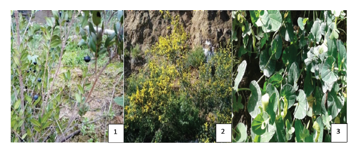
Medicinal plants Myrtus communis (L.), Calycotome spinosa (L.), and Aristolochia longa (L.) were collected in many areas in the littoral of Algeria (March 2018), namely Damous (Tipaza), Benni Haoua (Chlef), and Bissa (Chlef) (Fig. 1). These species were identified by Dr. Belhacini, a teacher and researcher at Hassiba Benbouali University of Chlef (Algeria).
Preparation of aqueous extracts. For each species we used dried and powdered plant aerial parts according to traditional use in these areas, namely leaves for M. communis and A. longa and leaves and flowers for C. spinosa. Aqueous extracts were prepared by a decoction of the plant material. In particular, 10 g of the plant material was boiled with 100 mL of distilled water for 15 min and then the solution was filtered and dried at 39°C.
Phytochemical screening. Phytochemical tests were performed on 5% infusion to detect certain secondary metabolites according to Takaidza et al. and Behbahani et al. [11, 12].
Determination of total phenol contents. A mixture of 250 μL of Folin Ciocalteu phenol reagent, 50 μL of the sample, and 500 μL of 20% Na2CO3 was prepared. The volume was adjusted to 5 mL with distilled water while shaking vigorously. After 30 min incubation, absorbance was read at 765 nm. A calibration curve of gallic acid (0–1 mg/mL) was done in parallel. The results were expressed in mg of gallic acid equivalent/g of dry matter (mg EAG/g DM) [13].
Determination of total flavonoid contents. The flavonoid assay was performed according to the method of Hmid et al. [14]. 1 mL of each extract was mixed with 1 mL of 2% AlCl3. After 10 min incubation, absorbance was read at 430 nm. The flavonoid concentrations were calculated using a calibration curve established with quercetin (0–40 μg/mL) and expressed in mg of quercetin equivalent/g of dry matter (mg EQ/g DM).
Hemolytic activity determination. A phosphate buffered saline (PBS) solution with pH = 7.4 was prepared by mixing the following compounds in appropriate concentrations: Na2HPO4 (10 Mm), KH2PO4 (1.8 Mm), KCl (2.7 Mm), and NaCl (137 Mm) [15]. A concentration range for each extract (M. communis, C. spinose, and A. longa) was prepared by diluting in PBS: 25, 50, 100, and 200 mg/mL. An erythrocyte suspension was prepared from the blood of a healthy donor in a heparin tube. After centrifugation at 2400 rpm for 10 min, the plasma was removed and the pellet was washed twice with PBS and then filled up with the same volume of plasma removed. The erythrocyte suspension was diluted 20 times with PBS.
Erythrocyte hemolysis assay. The hemolytic effect test of the species studied was carried out according to the method described by Haddouchi et al. and Guo-Xiang and Zai-Qun [16, 17]. We mixed 2950 μL of the erythrocyte suspension with 50 μL of aqueous extract for each species in a hemolysis tube. The operation was repeated three times for each concentration. The tubes were incubated at 37°C for one hour. During this period, 500 μL of each test was taken every 15 min (in 15, 30, 45, and 60 min) and added to 1.5 mL of PBS and then centrifuged again at 2400 rpm for 10 min. The absorbance of the hemoglobin leak in the supernatant was read at 548 nm against a blank containing PBS. A negative control tube was prepared under the same experimental conditions, 2950 μL of the erythrocyte suspension and 50 μL of the PBS buffer solution. On the other hand, a total hemolysis tube was prepared containing 250 μL of the erythrocyte suspension and 4750 μL of distilled water. Each test was repeated three times. The hemolysis rate of various extracts was calculated as a percentage (%) of total hemolysis after 15, 30, 45, and 60 min of incubation, according to the following formula:
![]()
Statistical analysis. Statistical analysis was done by One Way ANOVA. The data obtained were analyzed using the student’s t-test. A P value less than 0.01 was considered statistically significant.
РЕЗУЛЬТАТЫ И ИХ ОБСУЖДЕНИЕ
Phytochemical screening. The phytochemical screening allowed us to highlight the presence of some secondary metabolites (saponosides, tannins, alkaloids, flavonoids, and anthocyanins). The phytochemical tests carried out on the infused flowers and leaves of the selected plants are shown in Table 1.
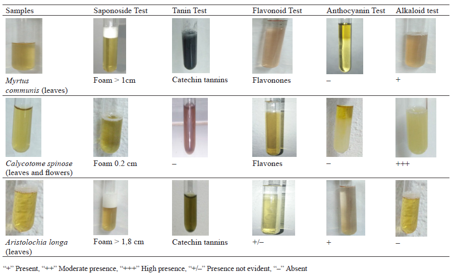
The results obtained after shaking the infusion for 15 min showed that Myrtus communis and Aristolochia longa were rich in saponosides because the foam was greater than 1 cm. In the Calycotome spinosa leave and flower infusion, the foam was unstable in the order of a few mm. The appearance of the orange and pink color after the addition of isoamyl alcohol indicated the presence of flavones in the C. spinosa leave and flower infusion. The purplish pink color indicated the presence of flavonones in the leaves of M. communis. In the A. longa infusion, the result was negative. The precipitate in the C. spinosa and M. communis infusions, which were previously acidified with sulfuric acid, after adding some drops of the Mayer reagent indicated the presence of alkaloids. However, the test was negative for A. longa. The appearance of a pink and red coloration after adding ammonia to the HCl-infused A. longa and C. spinosa indicated the presence of anthocyanins. However, this secondary metabolite was absent in the M. communis leave infusion.
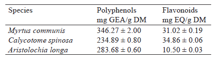
Contents of total polyphenols and flavonoids. The amount of polyphenols in the dry matter was expressed in mg gallic acid equivalent (mg EAG/g MS) and determined by the equation: y = 0.940x + b; R2 = 0.981. The a mount of flavonoids in the dry matter was expressed in mg of quercetin equivalent (mg EQ/g MS) and determined by the equation: y = 0.055x + b; R2 = 0.996 (Table 2). The total polyphenol content in the dry matter was 234.89 ± 0.80, 283.68 ± 0.60, and 346.27 ± 2.00 mg GEA/g for C. spinosa, A. longa, and M. communis, respectively. The content of total flavonoids in the dry matter was 10.50 ± 0.03, 34.86 ± 0.06, and 31.02 ± 0.19 mg EQ/g for A. longa, C. spinose, and M. communis, respectively (Table 2).
Hemolytic activity. In the negative control tube (tube containing only PBS and erythrocyte suspension), the hemolysis rate was constant and did not exceed 2.77 ± 0.35% after one hour of incubation. On the other hand, a total hemolysis of red blood cells was clearly observed in the total hemolysis tube. Indeed, we recorded a hemolysis rate that reached 99.86 ± 10.32% at 60 min.
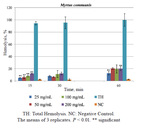
For the aqueous extract of the M. communis leaves, we observed a significantly low hemolysis rate during the first 15 min (P < 0.01). The hemolysis rates were 5.07 ± 0.21, 7.85 ± 1.20, and 12.57 ± 2.89% for the concentration of 25 mg/mL; 6.04 ± 1.90 (P < 0.01), 6.46 ± 0.77, and 20.42% for 50 mg/mL; 8.05 ± 1.41, 11.32 ± 5.72, and 19.51 ± 6.71 for 100 mg/mL; and 12.01 ± 0.21, 12.22 ± 0.26, and 20.14 ± 3.11% (P < 0.01) for 200 mg/mL, respectively, compared to total hemolysis (Fig. 2).
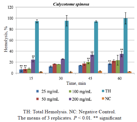
For C. spinosa, we found a significant increase in hemolysis rates over time (15, 30, 45, 60 min). Also, the rates were considerably higher with higher concentrations of the extract. For the concentrations of 25 and 50 mg/mL, hemolysis rates ranged between 6.52 ± 3.78 and 17.12 ± 1.50%, as well as 7.50 ± 2.95 and 22.36 ± 2.12%, respectively. However, a significant hemolytic effect was recorded in 100 (45 min) and 200 mg/mL (15, 45, and 60 min) of the C. spinosa extract. This rate increased from 8.14 ± 1.23% at 15 min to 23.61 ± 8.94% at 60 min in the presence of a 100 mg/mL concentration and from 24.44 ± 3.95% at 15 min to 34.86 ± 5.05% at 60 min in the presence of a 200 mg/mL concentration (Fig. 3).
For the extract of the A. longa leaves (Fig. 4), we found an increase in hemolysis rates over time (15, 30, 45, and 60 min). As the concentration increased, the percentage of hemolysis increased as well. At concentrations of 25 and 50 mg/mL, a hemolysis percentage ranged from 5 (15 min) to 6.71% (60 min) and from 7.22 (15 min) to 18.47% (60 min), respectively. The hemolysis rate was significant at 15 and 45 min (P < 0.01).
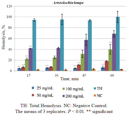
On the other hand, we observed an important hemolytic effect of the A. longa aqueous extract at concentrations of 100 and 200 mg/mL. This rate went from 22.12 ± 1.95 (15 min) to 40.23 ± 9.13% (60 min) and from 41.71 ± 0.75 (15 min) to 68.75 ± 6.11% (60 min), respectively. This increase in hemolytic effect remained inferior to total hemolysis.
Hemolytic effect of the plants studied at 60 min. Figure 5 shows the evolution of the hemolytic effect or the leakage of Hb after 60 min for the A. longa, C. spinosa, and M. communis extracts at four concentrations (25, 50, 100, and 200 mg/mL) in a PBS buffer medium (pH 7.4) containing an erythrocyte suspension incubated at 37°C, compared to a negative control tube (PBS + suspension) and a total hemolysis tube (distilled water + suspension).
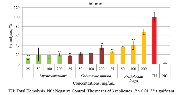
The M. communis species showed a significantly low hemoglobin leakage rate compared to the other species, as well as a 20.14 ± 3.11% total hemolysis. This species had a lesser effect on the cell membrane of erythrocytes (P < 0.01). However, C. spinosa caused a significant intermediate leakage of hemoglobin, compared to M. communis and A. longa, at 200 mg/mL (60 min), namely in the range of 34.86 ± 5.06% (P < 0.01). Nevertheless, the most important cytotoxic effect on red cells was produced by the aqueous extract of the A. longa leaves, where the leakage rate was 68.75 ± 6.11% at 200 mg/mL (60 min) and close to total hemolysis (P > 0.01), indicative of the species’ high toxicity. These results are phenotypically observable in the supernatant.
For millennia, humans have been searching for drugs in barks, seeds, fruit organs and other parts of plants to heal themselves and alleviate pain [18]. Nowadays, several studies have been conducted on plants to create new drugs and, to some extent, to evaluate their toxicity and identify their components. Polyphenolic compounds of M. communis L. extracts are grouped in three major chemical classes: phenolic acids, tannins, and flavonoids [19]. Our results of the phytochemical screening of C. spinosa are consistent with those reported by Cherfia et al., who identified polyphenols, flavonoids, alkaloids, tannins, and saponosides [20]. The Aristolochia species are a source of various active compounds such as aristolochic acid, alkaloids (aporphines, protoberberines, protopines), quinolines, amides, chlorophylls, terpenoids, lignans, flavonoids, tetralones, and steroids [21].
The polyphenol and flavonoid contents that we found in the M. communis leaves were higher than those obtained by Bouaziz et al., who reported 157.70 ± 2.65 mg EAG/g MS and 2.64 ± 0.22 mg EQ/g of dry matter [22]. In another study, the hydromethanolic extract of C. spinosa leaves had a polyphenol content of 228.42 ± 8.86 and a flavonoid content of 4.87 ± 0.12 [23]. According to Djeridane et al., the methanolic extract of A. longa contained 1.47 ± 0.20 mg/g EAG polyphenols and 0.81 ± 0.02 mg/g EQ flavonoids [24]. Our results were in agreement with Merouani et al., who found 396.88 ± 8.86 mg/g EAG polyphenols and 9.92 ± 0.23 mg/g EQ flavonoids [25].
Plants contain toxic compounds in high doses, which makes the evaluation of their hemolytic power indispensable for their correct use in traditional therapy, as well as for choosing the right mode of administration and preserving the integrity of membranes. According to Haddouchi et al., the hemolysis test should be performed even if a plant has a powerful antioxidant power, since its use in traditional medicine and in pharmacological preparations will be impossible in the presence of their hemolytic effect, which is an indicator of cytotoxicity [16]. “Free radicals induce several effects on erythrocytes, such as hemolysis, fluidity of the membrane, changes in morphometry and lipid peroxidation, among others. Erythrocytes potentially promoting the oxidative process are extremely sensitive to oxidative damage because of the polyunsaturated fatty acid content in their cell membranes and their high content of oxygen and hemoglobin” [26].
Many secondary metabolites were revealed in our extracts that may be a cause of cytotoxicity. We found a major lysis of red blood cells treated with C. spinosa, which was more prominent when treated with Aristolchia, testifying to severe toxicity. We found very few studies on A. longa and C. spinosa, trying to examine a relationship between the extracts’ chemical composition and toxicity.
According to Bissinger et al., saponins – a secondary metabolite identified in aqueous extracts of the plants under our study – may lead to the stimulation of hemolysis as well as to suicidal erythrocyte death [27]. Alkaloids are present in many plants which may be toxic and affect human health [28]. Mahdeb et al. reported that alkaloids are capable of disrupting the permeability of the membranes of erythrocytes [29].
As stated by Galati and O’Brien, many adverse effects were associated with dietary polyphenol consumption or exposures such as hemolytic anemia [30]. The authors added that before using these polyphenols for therapy, they need to be assessed for safety.
According to Grollman et al., the toxicity of Aristolochia longa is due to a toxin that is a major component of all Aristolochia species, namely the aristolochic acid responsible for nephropathic syndromes, although the therapeutic use of Aristolochia has rarely taken into account its intrinsic toxicity before [31]. These findings corroborate the study of Touiti et al., which showed that Aristolochia longa was incriminated in nephrotoxicity [32].
ВЫВОДЫ
Some herbs used in traditional therapy in high doses can reveal toxic properties and harm human health. It appears essential to determine their hemolytic capacity as a marker of toxicity for rational adaptation to traditional therapy. We found that Aristolochia longa and Calycotome spinosa caused significant lyses of red blood cells and a potent leakage of hemoglobin. Therefore, these species cannot be used without control as a therapeutic or pharmacological tool to treat diseases. Furthermore, it is important to perform antitumoral tests on cancer cells with these plant extracts or their chemical compounds to develop anticancer drugs.КОНФЛИКТ ИНТЕРЕСОВ
The authors declare no conflict of interest.СПИСОК ЛИТЕРАТУРЫ
- Atanasov AG, Waltenberger B, Pferschy-Wenzig E-M, Linder T, Wawrosch C, Uhrin P, et al. Discovery and resupply of pharmacologically active plant-derived natural products: A review. Biotechnology Advances. 2015;33(8):1582–1614. https://doi.org/10.1016/j.biotechadv.2015.08.001.
- Tembe FE, Pougoue KJ, Ngoupayo J, Njunkio BN, Nguidjoe E, Tabi YO, et al. Evaluation of the toxicity of secondary metabolites in aqueous extracts of Ficus thonningii Blume in Wistar rats. American Journal of Ethnomedicine. 2018;5(2).
- Calixto JB. Efficacy, safety, quality control, marketing and regulatory guidelines for herbal medicines (phytotherapeutic agents). Brazilian Journal of Medical and Biological Research. 2000;33(2):179–189. https://doi.org/10.1590/s0100-879x2000000200004.
- Kharchoufa L, Merrouni IA, Yamani A, Elachouri M. Profile on medicinal plants used by the people of North Eastern Morocco: Toxicity concerns. Toxicon. 2018;154:90–113. https://doi.org/10.1016/j.toxicon.2018.09.003.
- Forte JS, Raman A. Regulatory issues relating to herbal products – Part 2: Safety and toxicity. Journal of Medicinal Food. 2000;3(1):41–57. https://doi.org/10.1089/jmf.2000.3.41.
- Subramanian K, Sankaramourthy D, Gunasekaran M. Toxicity studies related to medicinal plants. In: Mandal SC, Mandal V, Konishi T, editors. Natural products and drug discovery: An integrated approach. Elsevier; 2018. pp. 491–505. https://doi.org/10.1016/b978-0-08-102081-4.00018-6.
- Quézel P, Santa S. Nouvelle flore de l’Algérie et des régions désertiques méridionales. Paris: Centre National de la Recherche scientifique; 1962. 1170 p.
- Bouzabata A, Casanova J, Bighelli A, Cavaleiro C, Salgueiro L, Tomi F. The genus Myrtus L. in Algeria: Composition and biological aspects of essential oils from M. communis and M. nivellei: A review. Chemistry and Biodiversity. 2016;13(6):672–680. https://doi.org/10.1002/cbdv.201500342.
- Alipour G, Dashti S, Hosseinzadeh H. Review of pharmacological effects of Myrtus communis L. and its active constituents. Phytotherapy Research. 2014;28(8):1125–1136. https://doi.org/10.1002/ptr.5122.
- Heinrich M, Chan J, Wanke S, Neinhuis W, Simmond MSJ. Local uses of Aristolochia species and content of nephrotoxic aristolochic acid 1 and 2 – A global assessment based on bibliographic sources. Journal of Ethnopharmacology. 2009;125(1):108–144. https://doi.org/10.1016/j.jep.2009.05.028.
- Takaidza S, Mtunzi F, Pilla M. Analysis of the phytochemical contents and antioxidant activities of crude extracts from Tulbaghia species. Journal of Traditional Chinese Medicine. 2018;38(2):272–279. https://doi.org/10.1016/j.jtcm.2018.04.005.
- Behbahani BA, Shahidi F, Yazdi FT, Mortazavi SA, Mohebbi M. Antioxidant activity and antimicrobial effect of tarragon (Artemisia dracunculus) extract and chemical composition of its essential oil. Journal of Food Measurement and Characterization. 2017;11(2):847–863. https://doi.org/10.1007/s11694-016-9456-3.
- Raafat K, Samy W. Amelioration of diabetes and painful diabetic neuropathy by Punica granatum L Extract and its spray dried biopolymeric dispersions. Evidence-based Complementary and Alternative Medicine. 2014;2014. https://doi.org/10.1155/2014/180495.
- Hmid I, Elothmani D, Hanine H, Oukabli A, Mehinagic E. Comparative study of phenolic compounds and their antioxidant attributes of eighteen pomegranate (Punica granatum L.) cultivars grown in Morocco. Arabian Journal of Chemistry. 2017;10:S2675–S2684. https://doi.org/10.1016/j.arabjc.2013.10.011.
- Mohan C. Buffers. A guide for the preparation and use of bufferd in biological systems. EMD Bioscience; 2006. 38 p.
- Haddouchi F, Chaouche TM, Halla N. Phytochemical screening, antioxidant activities and hemolytic power of four Saharan plants from Algeria. Phytotherapie. 2018;16(S1):S254–S262. https://doi.org/10.3166/phyto-2019-0140.
- Li G-X, Liu Z-Q. The protective effects of ginsenosides on human erythrocytes against hemin-induced hemolysis. Food and Chemical Toxicology. 2008;46(3):886–892. https://doi.org/10.1016/j.fct.2007.10.020.
- Petrovska BB. Historical review of medicinal plants usage. Pharmacognosy Reviews. 2012;6(11):1–5. https://doi.org/10.4103/0973-7847.95849.
- Aleksic V, Knezevic P. Antimicrobial and antioxidative activity of extracts and essential oils of Myrtus communis L. Microbiological Research. 2014;169(4):240–254. https://doi.org/10.1016/j.micres.2013.10.003.
- Cherfia R, Kara Ali M, Talhi I, Benaissa A, Kacem Chaouche N. Phytochemical analysis, antioxidant and antimicrobial activities of leaves and flowers ethyl acetate and n-butanol fractions from an Algerian endemic plant Calycotome spinosa (L.) Link. Journal of Pharmacognosy and Phytotherapy. 2017;9(12):185–196. https://doi.org/10.5897/JPP2017.0471.
- Tian-Shung W, Amooru GD, Chung-Ren S, Ping-Chung K. Chemical constituents and pharmacology of Aristolochi species. Studies in Natural Products Chemistry. 2005;32:855–1018. https://doi.org/10.1016/S1572-5995(05)80071-7.
- Bouaziz A, Khennouf S, Zarga MA, Abdalla S, Baghiani A, Charef N. Phytochemical analysis, hypotensive effect and antioxidant properties of Myrtus communis L. growing in Algeria. Asian Pacific Journal of Tropical Biomedicine. 2015;5(1):19–28. https://doi.org/10.1016/S2221-1691(15)30165-9.
- Krimat S, Dob T, Lamari L, Boumeridja S, Chelghoum C, Metidji H. Antioxidant and antimicrobial activities of selected medicinal plants from Algeria. Journal of Coastal Life Medicine. 2014;2(6):478–483.
- Djeridane A, Yousfi M, Nadjemi B, Vidal N, Lesgards JF, Stocker P. Screening of some Algerian medicinal plants for the phenolic compounds and their antioxidant activity. European Food Research and Technology. 2007;224(6):801–809. https://doi.org/10.1007/s00217-006-0361-6.
- Merouani N, Belhattab R, Sahli F. Evaluation of the biological activity of Aristolochia longa L. extracts. International Journal of Pharmaceutical Sciences and Research. 2017;8(5):1978–1992. https://doi.org/10.13040/IJPSR.0975-8232.8(5).1978-92.
- Martínez V, Mitjans M, Vinardell MP. Cytoprotective effects of polyphenols against oxidative damage. In: Watson RR, Preedy VR, Zibadi S, editors. Polyphenols in human health and disease. Vol. 1. Academic Press; 2014. pp. 275–288. https://doi.org/10.1016/B978-0-12-398456-2.00022-0.
- Bissinger R, Modicano P, Alzoubi K, Honisch S, Faggio C, Abed M, et al. Effect of saponin on erythrocytes. International Journal of Hematology. 2014;100(1):51–59. https://doi.org/10.1007/s12185-014-1605-z.
- Beyer J, Drummer OH, Maurer HH. Analysis of toxic alkaloids in body samples. Forensic Science International. 2009; 185(1–3):1–9. https://doi.org/10.1016/j.forsciint.2008.12.006.
- Mahdeb N, Mayouf S, Boukhari F, Souilah S, Bouzidi A. Hemolytic effect of total alkaloids from the seeds of Peganum harmala in vitro on erythrocytes of ruminants: Sheep, cattle and goats. Asian Journal of Plant Science and Research. 2013;3(6):53–59.
- Galati G, O'Brien PJ. Potential toxicity of flavonoids and other dietary phenolics: significance for their chemopreventive and anticancer properties. Free Radical Biology and Medicine. 2004;37(3):287–303. https://doi.org/10.1016/j.freeradbiomed.2004.04.034.
- Grollman AP, Scarborough J, Jelaković B. Aristolochic acid nephropathy: An environmental and iatrogenic disease. Advances in Molecular Toxicology. 2009;3:211–227. https://doi.org/10.1016/S1872-0854(09)00007-1.
- Touiti N, Achour S, Iken I, Chebaibi M, Houssaini TS. Nephrotoxicity associated with herbal medicine use, experience from Morroco. Toxicologie Analytique et Clinique. 2019;31(3):145–152. https://doi.org/10.1016/j.toxac.2019.04.001.
