

Аннотация
Introduction. New natural antioxidants remain a relevant research task of food science. Natural antioxidants neutralize free radicals in food systems, as well as in human body. The antioxidant properties of seaweed have attracted scientific attention for many years. However, most experiments featured non-polar extracts while aqueous extracts still remain understudied. The present research objective was to evaluate the antioxidant properties of hydrothermal extracts of edible seaweed from the Northern Coast of the Sea of Japan.Study objects and methods. The study featured hot-water and autoclave (30 and 60 min) extracts of three edible seaweed species from Russia’s Far East. The research focused on dry matter yield, total phenol content, phenolic profile, antiradical properties, hydroxylion (OH•) scavenging activity, and superoxide radical (O2•−) scavenging activity.
Results and discussion. The hot-water extracts appeared to have a higher yield than the autoclave extracts. The hot-water extract of red-purple seaweed Gracilaria verrucosa had the highest yield – 15.90%. The extract of brown seaweed Sargassum miyabei demonstrated the highest total phenol content. The phenolic profile of the extracts revealed 10 compounds, syringic acid and epicatechin being the major ones. The radical scavenging activity of the extracts varied from 48.2 to 88.9%, the highest value was observed in the hot-water extract of S. miyabei. The autoclave S. miyabei extracts also had a high radical scavenging activity, which exceeded other samples by 5.0–13.3%. The hot-water (30 min) extract of G. verrucosa had the lowest antiradical activity. Hot-water and autoclave extracts of S. miyabei showed the best OH• scavenging activity. Only the samples of G. verrucosa demonstrated signs of superoxide radical scavenging.
Conclusion. The extracts of brown seaweed S. miyabei proved to be the most active. The hot-water and autoclave extracts had the highest total phenol content and the strongest DPPH and OH• inhibitory activity.
Ключевые слова
Marine plants, phenols, extract, antioxidant activity, radicals, Sea of JapanВВЕДЕНИЕ
Modern science associates many diseases with the destructive effect of oxidants, or free radicals. Peroxidation is the most grievous consequence of free radicals entering the cell. Lipid oxidation in biological membranes is a highly destructive process that triggers liver damage, carcinogenesis, and aging. Lipid peroxidation is a major cause of the defective cell proliferation [1, 2]. Oxidized lipid compounds react with proteins, thus lowering enzymatic activity, causing cleavage of polypeptide chains, initiating DNA damage, accelerating aging, and aggravating such diseases as cancer and atherosclerosis. The aldehyde groups of these compounds form intermolecular crosslinks, which violate the structure of macromolecules and disorganize their functioning. By oxidizing lipids, free radicals cause glaucoma, cataracts, cirrhosis, ischemia, etc. [3]. The negative effects of oxidative stress can be mitigated by antioxidants, which neutralize potentially harmful reactive free radicals in the body cells before they cause lipid and protein oxidation. As a result, they can reduce potential mutations and prevent cancer and heart disease [4, 5].
Plants contain effective antioxidant substances. Polyphenols or bioflavonoids of plant origin, when they work in tandem, are extremely efficient against free radicals. Therefore, any study of vegetables, fruits, and spices as an alternative source of antioxidants will be highly relevant, considering that synthetic antioxidants are potentially toxic and carcinogenic. The past two decades have seen a lot of studies in phytochemicals from various types of terrestrial plant materials [6–8].
In this respect, seaweed attracts much less scientific attention than higher plants. Seaweed makes up a significant part of the marine flora. It contains compounds of various structures and bioactivities, which provide antioxidant, antibacterial, antiinflammatory, and anticarcinogenic properties [9–11]. Although seaweed is rich in polysaccharides and minerals, it is seldom used in food technology. A science-based proof of its antioxidant activity could increase its value as a food or supplement and expand its market. When exposed to light and oxygen, seaweed produces free radicals and other strong oxidants. However, the absence of oxidative damage in its structural components and its resistance to oxidation during storage suggest that seaweed has a good antioxidant system. Seaweed extracts are known to be rich in such hydrophilic components as polyphenols and soluble polysaccharides [9, 12–15]. In particular, brown seaweed is rich in natural antioxidants, e.g. such phenolic compounds as phlorotannins and such carotenoids as fucoxanthin and isoprenoids [16].
Many seaweed extracts possess strong antioxidant properties [17–21]. However, there have been no publications on the antioxidant activity of seaweed from the Northern Coast of the Sea of Japan. Moreover, the antioxidant activity of seaweed has been studied mainly in non-polar extracts and very seldom – in aqueous extracts [22, 23]. Seaweed owes its antioxidant properties due to its hydrophilic and hydrophobic compounds. Therefore, its antioxidant activity in hydrothermal extracts needs more research because hydrothermal treatment is an integral part of processing.
This study featured three species of seaweeds that are part of human and animal diet in the Primorye Territory of Russia’s Far East.
Codium flagile from the family of Codiaceae is a species of green seaweed found in the Indo-Pacific and the Atlantic. In Russia, it grows on the Northern Coast of the Primorye Territory. Its thallus is prostrate, branched, pillow-shaped or upright. It can be 5–30 cm long. In Oceania and Asia, and especially in Korea and Russia’s Primorye, C. flagile is usually served raw or boiled. C. flagile extracts are widely used in medical cosmetology as it synthesizes water-retaining substances.
Gracilaria verrucosa, Gracilariaceae family, is a species of red-purple seaweed found in the lowboreal-tropical Atlantic-Indo-Pacific. In Russia, this seaweed grows in the Far East, in the Sea of Japan and the Sea of Okhotsk. It has branched, cylindrical or flat, gristly thallus, which is purple-red or fading to green. G. verrucosa is 20–40 cm long, sometimes reaching up to 100 cm. It grows in the littoral and sublittoral sea zones at a depth of up to 2 m. The species prefers estuarine and sheltered coastal areas and stony, siltysandy and sandy bottom, interspersed with stones and shell rock. Classified as a potentially commercial species, G. verrucosa is a fast-growing seaweed with a biomass of 4.5 kg/m2, which makes it convenient for cultivation. In Southeast Asia, it is used for food and as a tonic substance in folk medicine. G. verrucosa can serve as a raw material for agar and various food and feed additives. It is known to contain minerals, proteins with antibiotic properties, B vitamins, phycocolloids, etc. However, agar remains the only commercial product related to this raw material.
Sargassum miyabei, Sargassaceae (Decne) Kutz. family, is a species of brown seaweed that thrives in cold waters in the low-boreal-subtropical areas of Asian. Its thallus is branched, rough, and thick, 2.0–2.5 m in length, olive green. It grows in the littoral and sublittoral at a depth of 11 m on rocky, stony, and silty-sandy bottom, near semi-sheltered and open coastal areas, in separate bushes or small clusters, clustering with kelp seaweeds and sea grasses. It is a potentially commercial perennial species, with the biomass up to 15 kg/m2, the mass of one thallus up to 7 kg, and population density of 2–6 specimens per 2 m2. In the Russian Far East, it grows in the Sea of Japan and the Sea of Okhotsk. It is used in the food industry and as a raw material for therapeutic and prophylactic medications, food and feed additives. S. miyabei is known to contain vitamins A, В1, В6, B12, minerals, polysaccharides with immunostimulating and antitumor activity, etc. [24].
The research objective was a comparative analysis of hydrothermal extracts of the following seaweeds: green C. flagile, brown S. miyabei, and red G. verrucosa, obtained by hot-water or autoclave methods for 30 and 60 min. The extracts were compared according to the following parameters: total phenols, phenolic profile, and antioxidant activity, i.e. DHHP radicals, OH• scavenging activity, and O2•− scavenging activity.
ОБЪЕКТЫ И МЕТОДЫ ИССЛЕДОВАНИЯ
Materials. All samples of Codium flagile, Gracilaria verrucosa, and Sargassum miyabei were harvested in the coastal sea waters of the Sea of Japan in the northern Primorye Territory in May-July 2018 (Table 1).
Chemicals and reagents. 1,1-diphenyl-2-picrylhydrazyl (DHHP), ВНТ-2,6-di-tert-butyl-4-methylphenol (ionol), and tannic acid were purchased from Sigma-Aldrich (USA). The Folin-Ciocalteu phenolic reagent was purchased from Fluka (Switzerland). All the other reagents were of analytical grade.
Extract samples. After harvesting, the samples were placed in plastic bags, cooled with ice, and immediately transported to the laboratory. They were washed with flowing tap water to remove salt, sand, and epiphytes. After that, the samples were soaked twice in distilled water for 30 min, dried on filter paper, and ground using a Waring mill. The samples were stored in an airtight container at –20°C. Hot water extraction procedures followed the methods described in [25, 26]. To obtain hot-water extracts, 10 g of crushed seaweed was added to 300 mL of deionized water and boiled for 30 and 60 min. Then, the suspension was filtered through three layers of gauze. The resulting filtrate underwent freezedrying and subsequent grinding to obtain a dry extract. To obtain autoclave extracts, 10 g of crushed seaweed was autoclaved in 100 mL of deionized water at 120°C for 30 and 60 min. The hot aqueous solution was filtered through three layers of gauze. The residue was freezedried and crushed.
Total phenol content. A spectrophotometric method with the Folin-Ciokalteu reagent was used to determine the total content of phenolic compounds: a mix of phosphotungstic and phosphoromolybdic acids was reduced in an alkaline medium. It is the main method for determining the total content of phenols in medicine and food industry [27]. The experiment featured a UV-1800 scanning spectrophotometer (Shimadzu, Kyoto, Japan). The quantity of polyphenolic compounds was determined in terms of tannic acid.
Phenolic compounds were identified by HPLC in a high pressure liquid chromatograph LC-20A (Shimadzu, Kyoto, Japan) at 30°С on a reverse phase column (Phenomenex RPC18 250×4.6 mm, 5 μm, Torrance, California, USA). The extracts went through a 0.45 μm filter (Millipore, Westboro, MA, USA) before being put into the chromatograph. The total run time was about 50 min at a flow rate of 0.6 mL/min. The mobile phase included methanol (B) with water (A) and 0.2% acetic acid (B:A = 65:35, w/w). The gradient elution looked as follows: 0–2 min, 5% of isocratic B; 2–10 min, linear gradient 5–25% of B; 10–20 min, linear gradient 25–40% of B; 20–30 min, linear gradient 40–50% of B; 30–40 min, linear gradient 50–100% of B, 40–45 min, 100% of isocratic B, 45–55 min, linear gradient 100% of B. Individual phenolic compounds were identified by comparing retention times of phenolic compounds and original standards (Sigma, USA) under the same conditions. The simultaneous detection wavelength control was 324 nm for chlorine, caffeic, 2,5-dihydroxybenzoic, coumaric, ferulic, and salicylic acids and 277 nm for epigalocatechin gallate, epicatechin, epicatechin gallate, and syringic acid. Each compound was quantified based on its peak area.
Antioxidant activity. The antiradical properties of the extracts were assessed by their ability to interact with a stable free 2,2-diphenyl-1-picrylhydrazyl (DPPH) radical in vitro. The reaction mix contained 3 mL of 0.3 mM DPPH in ethanol, 1 mL of 50 mMtris-HCl buffer (pH 7.4), and 1 mL of extract [28].
After a 30-min incubation at room temperature, the optical density values were recorded at λ = 517 nm using a UV-1800 scanning spectrophotometer (Shimadzu, Japan) in 1×1 cm cuvettes at 25°C.
The radical scavenging properties were described using the following indicators: radical scavenging activity; the effective concentration of the substance which restores 50% of DPPH (ЕС50), mg/mL; recovery time for one half of the radical (TЕС50), min; and antiradical effectiveness.
The radical scavenging activity (RSA) was calculated as follows:
![]()
where A0 is optical density of the control solution and A1 is optical density of the extract.
Antiradical efficacy (AE) links the half-recovery time of the radical (TЕС50) and the required substrate concentration (ЕС50), which was calculated by the formula:
![]()
The antiradical properties were compared with the effect of the synthetic antioxidant ionol (2,6-ditert-butyl-4-methyl-phenol), which was preliminarily recrystallized from ethanol. Tthe obtained crystals were dried and sublimated in vacuum.
OH• scavenging activity. The OH• scavenging activity was determined according to [29]: 1.5 mM FeSO4 (0.5 mL) was mixed with 6 mM H2O2 (0.35 mL), 20 mM sodium salicylate (0.15 mL), and various concentrations (0.2–1.0 mg/mL) of 1 mL of each sample. The mix was kept at 37°C for 1 h. The optical density of the hydroxylated salicylate complex was determined spectrophotometrically at a wavelength of 562 nm. Ascorbic acid was used as positive control. The OH• scavenging activity was calculated using the following formula:
![]()
where A1 is optical density of the extract; A0 is optical density of the control solution; and A2 is optical density of the reagent blank without sodium salicylate.
O2•− scavenging activity. The O2•− scavenging activity was determined according to the method developed by Ruch et al. [30]: 40 mM Н2О2 was dissolved in phosphate buffer (pH 7.4). Various concentrations (0.1–1.0 mg/mL) of extracts in MeOH (3.0 mL) were added to a 40 mM H2O2 solution (3.0 mL). The optical density was measured at 230 nm after 10 min of incubation against an empty solution of phosphate buffer without H2O2.
The O2•− scavenging activity was determined by the formula:
![]()
where A1 is optical density of the extract and A0 is optical density of the control solution.
Calculation of ЕС50 (mg/mL) was the next step.
Statistical analysis. The data were obtained as mean and standard deviation (SD) and analyzed by one-way ANOVA using SPSS 11.5 for Windows. The difference in mean values was significant at P < 0.05.
РЕЗУЛЬТАТЫ И ИХ ОБСУЖДЕНИЕ
The hot-water and autoclave hydrothermal extracts obtained from three edible seaweeds from the Northern Coast of the Sea of Japan demonstrated different dry matter yields. Figure 1 shows the effect of processing time and method on the extraction yield.
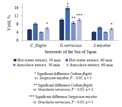
A comparative analysis of the quantitative yield resulted in the following descending order: Gracilaria verrucosa > Codium flagile > Sargassum miyabei.
The hot-water extract of the red-purple seaweed G. verrucosa had the highest yield – 15.90%, which was 1.17–1.58 times higher than that in the autoclave samples. The extraction time increased the yield by 55–57% for hot-water extracts and by 16.3–47.5% for autoclave extracts. The hot-water G. verrucosa extract and the autoclave C. flagile and S. miyabei extracts experienced the greatest effect of extraction time. However, a high yield did not equal high antioxidant activity, which depends on strong antioxidants, e.g. polyphenols.
Figure 2 illustrates the content of phenols in the seaweed extracts.
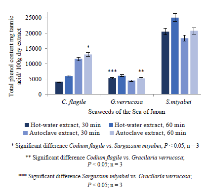
A comparative analysis of the total phenols showed that S. miyabei had the hot-water extract with the highest phenolic level, about 3–4 times higher than the other hot-water extracts. The autoclave extract of S. miyabei had similar level of total phenols. The extract of C. flagile had the lowest total phenols among the hot-water extracts, while G. verrucosa had the lowest total phenols among the autoclave samples. The fact that brown seaweeds proved richer in fenols than green and red seaweeds confirms the data obtained by other scientists [31]. However, the methods were slightly different. The total phenol content in the autoclave C. flagile extract was twice as high as in the hot-water extract, while in the autoclave extract of S. miyabei it was lower than in the respective hot-water extracts (Fig. 2).
Such hydrophilic polyphenols as phlorotanins, which are bipolar and occur in brown seaweeds, can act as antioxidant components and thus help the seaweed overcome oxidative stress [32, 33].
Identification of phenolic compounds. Individual phenolic compounds were tested using HPLC in extracts obtained after 60 min of hydrothermal treatment because a longer processing time provided a higher level of total phenols. Table 2 shows the content of the main phenolic compounds in the extracts.
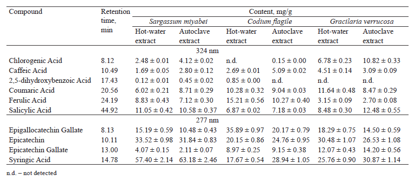
Table 2 demonstrates 10 phenolic compounds in the composition of edible seaweed extracts from the Northern Coast of the Sea of Japan. Chlorogenic acid was the first of the phenolic compounds to elute; it was not detected in the hot-water C. flagile extract. Similarly, 2,5-dihydroxybenzoic acid was not detected in both extracts of G. verrucosa and the autoclave extract of C. flagile. Caffeic, coumaric, ferulic, salicylic, and syringic acids, epigallocatechin gallate, epicatechin gallate, as well as epicatechin were registered in all the samples. Syringic acid and epicatechin appeared to be the major phenolic compounds for all the extracts.
All the autoclave extracts showed higher levels of chlorogenic and syringic acids compared to the hotwater samples, which had a higher content of ferulic acid and epigallocatechin gallate.
A comparative analysis of individual phenolic compounds showed that S. miyabei extracts had the highest content of epicatechin, which was maximal in the hot-water sample, and syringic acid, which was maximal in the autoclave sample. The C. flagile extracts demonstrated the highest content of ferulic acid and epigallocatechin gallate, which were the most abundant in the hot-water sample. The G. verrucosa extracts had the highest content of chlorogenic acid, especially in the autoclave sample, coumaric acid (hot-water sample), salicylic acid (autoclave sample), and epicatechin gallate (autoclave sample). Thus, the phenolic profile of the extracts depended on the type of seaweed and the method of hydrothermal treatment. Phenolic acids, which are the main class of phenolic compounds, are found in seaweed in significant quantities. Typical phenols are known to possess antioxidant properties. Other publications prove that seaweed is rich in phenolic compounds, e.g. catechin, caffeic acid, epicatechin, epicatechin gallate, etc. [34, 35].
Antioxidant activity. Radical scavenging properties. DPPH possesses a nitrogen free radical and is easily destroyed by a free radical scavenger. The DPPH radical assay was used to test the ability of the antioxidant compounds found in the extracts to act as proton radical scavengers or hydrogen donors. Table 3 demonstrates the antiradical properties of the extracts.
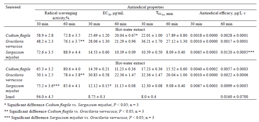
The autoclave extracts appeared to be more active than the hot-water extracts. The radical scavenging activity varied within wide limits of 39.6–88.9%. The maximal value was observed in the hot-water S. miyabei extract, and its radical scavenging activity was 12.8–28.4% higher than that of the other extracts and only 10.4% lower than that of ionol. The autoclave extracts of S. miyabei also had a high radical scavenging activity, which exceeded that of the other extracts by 5.0–13.3%. A comprehensive assessment of the antiradical activity showed that the hot-water (30 min) extract of G. verrucosa had the lowest values. The hot-water extract of S. miyabei had the most pronounced antiradical properties. Its ЕС50 concentration was 1.89–2.41 times lower than the other extracts, its time was 2.21–9.79 times shorter, and its antiradical efficiency was 4.29–24.00 times higher.
The extract of S. miyabei, which had the highest total phenol content, also had a much lower effective concentration of ЕС50. The autoclave extracts demonstrated a strong correlation between the level of total phenols (Fig. 2) and the effective concentration of ЕС50. When the total phenol level was high, the effective ЕС50 concentration was low. Therefore, the polyphenolic components in the seaweed extracts were capable of acting as free radical scavengers. For hot-water extracts, however, this ratio was weak. Since the DPPH radical analysis was not specific for any particular antioxidants, the antiradical activity of these extracts result not only from phenols, but also from various other water-soluble antioxidants, e.g. polysaccharides, folic acid, thiamine, and ascorbic acid [36, 37].
The OH• scavenging activity and absorption of superoxide radicals. Antioxidant activity was determined according to the ability of the antioxidant components to inhibit the oxidation of deoxyribose by the reactive hydroxyl-ion radical (OH•), formed as a result of the Fenton-type reaction. The reaction involves two antioxidant defense mechanisms: suppression of the generation of OH• from H2O2 by binding with metal ions and the direct transfer of one electron to the generated radical. Figure 3 illustrates the OH• scavenging activity of the seaweed extracts.
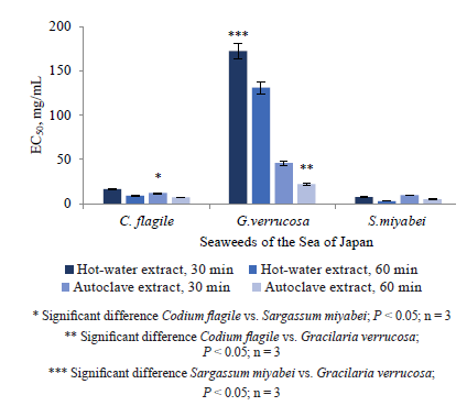
Both autoclave and hot-water S. miyabei extracts demonstrated the highest OH• scavenging activity, while that of the other extracts was significantly lower. A comparative analysis showed similar ЕС50 values and ranking. A longer extraction time increased the OH• scavenging activity, but the increase was insignificant. Brown seaweeds are known to contain floratanins, which chelate heavy metals [32, 38]. Probably, the high content of floratanins predetermined the high OH• scavenging activity of S. miyabei extracts. However, seaweeds have a complex composition, and its phenols are not the only compounds that exhibit antioxidant activity, which is also typical of carotenoids and polysaccharides [39]. Hydrothermal extracts of some green and brown seaweeds contain sulfated heteropolysaccharides, which strongly inhibit OH• activity in vitro [40–42].
Figure 4 demonstrates the O2•− scavenging activity of the extracts.
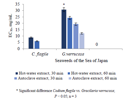
Although O2•− is a relatively weak oxidizing agent, it decomposes into stronger oxidative forms, such as singlet oxygen and OH•. The autoclave extract of G. verrucosa was the only sample to demonstrate O2•− scavenging activity. All hot-water extracts showed O2•− inhibitory activity. Figure 4 shows that the maximal inhibitory activity belonged to the C. flagile extract. Surprisingly, S. miyabei revealed no signs of O2•− scavenging. The extraction time only slightly increased the O2•− absorption.
The established difference in the O2•− absorption between the hot-water and autoclave extracts proved that the two methods extracted different types of antioxidants. The lack of correlation between the total content of phenols and O2•− inhibition means that such activity demonstrated by all three species could not be explained solely by the presence of phenolic antioxidants. Sulfated polysaccharides obtained from the hot-water extracts of green and brown seaweeds inhibited the activity of O2•− in vitro [40–42].
ВЫВОДЫ
The hot-water extraction proved more effective than the autoclave extraction in terms of OH• and O2•− scavenging activity. However, the autoclave extracts had better antiradical properties. The extracts of the brown seaweed Sargassum miyabei showed significantly higher antiradical and antioxidant activity than the extracts of the red seaweed Gracilaria verrucosa and the green seaweed Codium flagile. The relationship between OH• scavenging and antioxidant activities in these samples indicated that it came from the hydrophilic polyphenolic antioxidants. The hydrothermal extracts of the seaweeds proved to be a promising source of antioxidants for human consumption.КОНФЛИКТ ИНТЕРЕСОВ
The authors declare that there is no conflict of interests related to the publication of this article.ФИНАНСИРОВАНИЕ
The research was supported by the Presidential grant No. МК-4715.2021.4.СПИСОК ЛИТЕРАТУРЫ
- Tan BL, Norhaizan ME. Liew W-P-P. Nutrients and oxidative stress: Friend or foe? Oxidative Medicine and Cellular Longevity. 2018;2018. https://doi.org/10.1155/2018/9719584.
- Sies H. Hydrogen peroxide as a central redox signaling molecule in physiological oxidative stress: Oxidative eustress. Redox Biology. 2017;11:613–619. https://doi.org/10.1016/j.redox.2016.12.035.
- Mazon JN, de Mello AH, Ferreira G, Rezin GT. The impact of obesity on neurodegenerative diseases. Life Sciences. 2017;182:22–28. https://doi.org/10.1016/j.lfs.2017.06.002.
- Noufou O, Sawadogo WR, Tibiri A, Lompo M, Dijoux MG, Diederich M, et al. Phenolic contents and in vitro pharmacological activities of methanolic extract of Pterocarpus erinaceus Poir. Stem bark (Fabaceae). Journal of Pharmaceutical Research International. 2016;10(2). https://doi.org/10.9734/BJPR/2016/23149.
- Halliwell B, Aeschbach R, Löliger J, Aruoma OI. The characterization of antioxidants. Food and Chemical Toxicology. 1995;33(7):601–617. https://doi.org/10.1016/0278-6915(95)00024-V.
- Wang S, Zhu F. Chemical composition and biological activity of staghorn sumac (Rhus typhina). Food Chemistry. 2017,237: 431–443. https://doi.org/10.1016/j.foodchem.2017.05.111.
- Li Y, Jiang J-G. Health functions and structure-activity relationships of natural anthraquinones from plants. Food and Function. 2018;9(12):6063–6080. https://doi.org/10.1039/c8fo01569d.
- Fierascu RC, Ortan A, Fierascu IC, Fierascu I. In vitro and in vivo evaluation of antioxidant properties of wild-growing plants. A short review. Current Opinion in Food Science. 2018;24:1–8. https://doi.org/10.1016/j.cofs.2018.08.006.
- Meresse S, Fodil M, Fleury F, Chenais B. Fucoxanthin, a marine-derived carotenoid from brown seaweeds and microalgae: A promising bioactive compound for cancer therapy. International Journal of Molecular Sciences. 2020;21(23). https://doi.org/10.3390/ijms21239273.
- Jesumani V, Du H, Aslam M, Pei P, Huang N. Potential use of seaweed bioactive compounds in skincare – a review. Marine Drugs. 2019;17(12). https://doi.org/10.3390/md17120688.
- Gerasimenko NI, Martyyas EA, Busarova NG. Composition of lipids and biological activity of lipids and photosynthetic pigments from algae of the families Laminariaceae and Alariaceae. Chemistry of Natural Compounds. 2012;48(5):737–741. https://doi.org/10.1007/s10600-012-0371-5.
- Liu Z. Gao T, Yang Y, Meng F, Zhan F, Jiang Q, et al. Anti-cancer activity of porphyran and carrageenan from red seaweed. Molecules. 2019;24(23). https://doi.org/10.3390/molecules24234286.
- Salehi B, Sharifi-Rad J, Seca AML, Pinto DCGA, Michalak I, Trincone A, et al. Current trends on seaweeds: Looking at chemical composition, phytopharmacology, and cosmetic applications. Molecules. 2019;24(22). https://doi.org/10.3390/molecules24224182.
- Amosova AS, Ivakhnov AD, Skrebets TE, Ulyanovskiy NV, Bogolitsyn KG. Supercritical fluid extraction of carotenoids from Shantane carrot. Russian Journal of Physical Chemistry B. 2014;8(7):963–966. https://doi.org/10.1134/S1990793114070021.
- Rosa GR, Barreto MC, Seca AML. Pharmacological effects of Fucus spiralis extracts and phycochemicals: A comprehensive review. Botanica Marina. 2019;62(2):167–178. https://doi.org/10.1515/bot-2018-0047.
- Sukhoveeva MV, Podkorytova AV. Promyslovye vodorosli i travy morey Dalʹnego Vostoka: biologiya, rasprostranenie, zapasy, tekhnologiya pererabotki [Commercial seaweeds and sea grasses of the Far East: biology, distribution, reserves, and processing technology]. Vladivostok: TINRO-Tsentr; 2006. 243 p. (In Russ.).
- Cao L, Lee SG, Lim KT, Kim H-R. Potential anti-aging substances derived from seaweeds. Marine drugs. 2020;18(11). https://doi.org/10.3390/md18110564.
- Hentati F, Tounsi L, Djomdi D, Pierre G, Delattre C, Ursu AV. Bioactive polysaccharides from seaweeds. Molecules. 2020;25(14). https://doi.org/10.3390/molecules25143152.
- Viji P, Venkateshwarlu G, Ravishankar CN, Gopal TKS. Role of plant extracts as natural additives in fish and fish products – A review. Fishery Technology. 2017;54(3):145–154.
- Oucif H, Benaissa M, Ali Mehidi S, Prego R, Aubourg SP, Abi-Ayad S-ME-A. Chemical composition and nutritional value of different seaweeds from the west Algerian coast. Journal of Aquatic Food Product Technology. 2020;29(1):90–104. https://doi.org/10.1080/10498850.2019.1695305.
- Rajauria G. In-vitro antioxidant properties of lipophilic antioxidant compounds from 3 brown seaweed. Antioxidants. 2019;8(12). https://doi.org/10.3390/antiox8120596.
- Chakraborty K, Joseph D. Antioxidant potential and phenolic compounds of brown seaweeds Turbinaria conoides and Turbinaria ornata (class: Phaeophyceae). Journal of Aquatic Food Product Technology. 2016;25(8):1249–1265. https://doi.org/10.1080/10498850.2015.1054540.
- Chakraborty K, Maneesh A, Makkar F. Antioxidant activity of brown seaweeds. Journal of Aquatic Food Product Technology. 2017;26(4):406–419. https://doi.org/10.1080/10498850.2016.1201711.
- Dzizyurov VD, Kulepanov VN, Shaposhnikova TV, Sukhoveeva MV, Gusarova IS, Ivanova NV. Atlas massovykh vidov vodorosley i morskikh trav rossiyskogo Dalʹnego Vostoka [Atlas of mass species of seaweeds and sea grasses of the Russian Far East]. Vladivostok: TINRO-Tsentr; 2008. 328 p. (In Russ.).
- Hou W-Y, Chen J-C. The immunostimulatory effect of hot-water extract of Gracilaria tenuistipitata on the white shrimp Litopenaeus vannamei and its resistance against Vibrio alginolyticus. Fish and Shellfish Immunology. 2005;19(2):127–138. https://doi.org/10.1016/j.fsi.2004.11.009.
- Huang R, Lee H-T. Immunological properties of the marine brown alga Endarachne binghamiae (Phaeophyceae). International Journal of Applied Science and Engineering. 2005;3(3):167–173.
- FFS 1.5.3.0008.15. The determination of tannins in herbal drugs and medicinal plant preparations [Internet]. [cited 2020 Dec 20]. Available from: https://193.232.7.120/feml/clinical_ref/pharmacopoeia_2/HTML/#417/z.
- Molyneux P. The use of the stable free radical diphenylpicrylhydrazyl (DPPH) for estimating antioxidant activity. Songklanakarin Journal of Science and Technology. 2004;26(2):211–219.
- Smirnoff N, Cumbes QJ. Hydroxyl radical scavenging activity of compatible solutes. Phytochemistry. 1989;28(4):1057–1060. https://doi.org/10.1016/0031-9422(89)80182-7.
- Ruch RJ, Cheng S-J, and Klaunig E. Prevention of cytotoxicity and inhibition of intercellular communication by antioxidant catechins isolated from Chinese green tea. Carcinogenesis. 1989;10(6):1003–1008. https://doi.org/10.1093/carcin/10.6.1003.
- Garicano Vilar E, O’Sullivan MG, Kerry JP, Kilcawley KN. Volatile compounds of six species of edible seaweed: A review. Algal Research. 2020;45. https://doi.org/10.1016/j.algal.2019.101740.
- Papitha R, Selvaraj CI, Palanichamy V, Arunachalam P, Roopan SM. In vitro antioxidant and cytotoxic capacity of Kappaphycus alvarezii successive extracts. Current Science. 2020;119(5):790–798.
- Phenolic content and antioxidant capacity in algal food products Sathya R, Kanaga N, Sankar P, Jeeva S. Antioxidant properties of phlorotannins from brown seaweed Cystoseira trinodis (Forsskal) C. Agardh. Arabian Journal of Chemistry. 2017;10:S2608–S2614. https://doi.org/10.1016/j.arabjc.2013.09.039.
- Alkhalaf MI. Chemical composition, antioxidant, anti-inflammatory and cytotoxic effects of Chondrus crispus species of red algae collected from the Red Sea along the shores of Jeddah city. Journal of King Saud University Science. 2021;33(1). https://doi.org/10.1016/j.jksus.2020.10.007.
- Machu L, Misurcova L, Ambrozova JV, Orsavova J, Mlcek J, Sochor J, et al. Phenolic content and antioxidant capacity in algal food products. Molecules. 2015;20(1):1118–1133. https://doi.org/10.3390/molecules20011118.
- Wang L, Oh JY, Hwang J, Ko JY, Jeon Y-J, Ryu B. In vitro and in vivo antioxidant activities of polysaccharides isolated from celluclast-assisted extract of an edible brown seaweed, Sargassum fulvellum. Antioxidants. 2019;8(10). https://doi.org/10.3390/antiox8100493.
- Gomez-Zavaglia A, Prieto Lage MA, Jimenez-Lopez C, Mejuto JC, Simal-Gandara J. The potential of seaweeds as a source of functional ingredients of prebiotic and antioxidant value. Antioxidants. 2019;8(9). https://doi.org/10.3390/antiox8090406.
- Valdes FA, Gabriela Lobos M, Diaz P, Saez CA. Metal assessment and cellular accumulation dynamics in the green macroalga Ulva lactuca. Journal of Applied Phycology. 2018;30(1):663–671. https://doi.org/10.1007/s10811-017-1244-x.
- Tabakaeva OV, Tabakaev AV. Carotenoid profile and antiradical properties of brown seaweed Sargassum miyabei extracts. Chemistry of Natural Compounds. 2019;55(2):364–366. https://doi.org/10.1007/s10600-019-02692-w.
- Jridi M, Mezhoudi M, Abdelhedi O, Boughriba S, Elfalleh W, Souissi N, et al. Bioactive potential and structural characterization of sulfated polysaccharides from Bullet tuna (Auxis Rochei) by-products. Carbohydrate Polymers. 2018;194:319–327. https://doi.org/10.1016/j.carbpol.2018.04.038.
- Bogolitsyn K, Dobrodeeva L, Druzhinina A, Ovchinnikov D, Parshina A, Shulgina E. Biological activity of a polyphenolic complex of Arctic brown algae. Journal of Applied Phycology. 2019;31(5):3341–3348. https://doi.org/10.1007/s10811-019-01840-7.
- Layana P, Xavier KAM, Lekshmi S, Deshmukhe G, Nayak BB, Balange AK. Antioxidant and antimicrobial potential of hydroethanolic extracts of Padina tetrastromatica from North-west Coast of India. Fishery Technology. 2019;56(3):199–204.
