

Аннотация
Introduction. Microorganisms of dairy raw materials tend to adhere to the surfaces of processing equipment and form sustainable biofilms, which is a serious issue in the dairy industry. The goal of the present work was to investigate formation of biofilms on a glass surface in static model conditions, and removal of such biofilms by cleaning.Study objects and methods. The study objects were the permeates of skim milk, sweet whey and acid whey, as well as the biofilms formed and washings from glass slides. Biofilms were removed from the glass with detergents used in the dairy industry. Standard methods of determining microbiological and physicochemical properties were used to characterize the permeates. The biofilm structure and morphology of microorganisms participating in biofilm formation were investigated with a light spectroscopy. The efficiency of biofilm removal in a cleaning process was quantified with optical density of washings.
Results and discussion. Biofilms in whey permeates formed slower compared to those in skimmed milk permeate during the first 24 h. Yeasts contributed significantly to the biofilm microflora in acid whey permeate throughout 5 days of biofilm growth. Well adhered biofilm layers were the most stable in skimmed milk permeate. The highest growth of both well and poorly adhered biofilm layers was observed in sweet whey permeate after 3–5 days. It was established that the primary attachment of microorganisms to a glass surface occurred within 8 h, mature multicultural biofilms formed within 48 h, and their partial destruction occurred within 72 h.
Conclusion. The research results can be used to improve the cleaning equipment procedures in processing secondary dairy raw materials.
Ключевые слова
Biofilm, whey, formation, removal, filtration, dairy industry, secondary dairy raw materialsВВЕДЕНИЕ
Biofilms are complex microbial ecosystems formed at the interface usually on hard surfaces. Different types of viruses, bacteria and fungi in natural biofilms coexist, immersed in an extracellular matrix of polysaccharides, proteins and DNA. The matrix provides structural and functional benefits for biofilm microorganisms, including hydration, uptake and digestion of nutrients, protection from adverse environmental conditions, and exchange of genetic information [1].
Biofilms is a serious problem in the food industry as they can form on equipment and food surfaces. Biofilm microorganisms release substances that destroy the equipment material and spoil food products. The development of pathogenic microbes in biofilms is most dangerous, as they can acquire resistance to antimicrobial drugs in such communities.
Traditional chemical, physical and mechanical methods of removing contaminants and suppressing microorganisms used in the food industry are not always effective enough against biofilms. Therefore, different approaches are used to control them: proteases enzymatic treatment, glycosidases or DNAs; a steel surface modification by applying silver, copper or zinc nanoparticles, or new antibiofilm polymers with lysozyme or bacteriocins; introduction of biosurfactants into detergents, etc. [2, 3].
The biofilm formation in the equipment surface significantly affects the quality and safety of dairy products [4, 5]. The recent increase of biofilm research publications about the dairy industry indicates the relevance of this research (Fig. 1).

A biofilm goes through a number of stages in its development: an initial attachment, an irreversible adhesion, an early development of the structure (microcolonies formation), а growth of biofilm layers, а formation of a complex three-dimensional structure, and its subsequent partial destruction [2, 6]. During processing, the components of dairy raw materials can settle on the surface of the equipment, which creates favorable conditions for the fixation of microorganisms and the development of biofilms. Their structure and properties significantly depend on the type of processed raw material, its bacterial contamination, and processing conditions. The emerging communities of microorganisms surrounded by an exopolymer layer are very resistant to thermal and chemical treatment. Therefore, a biofilm formation reduces the efficiency of washing and disinfection procedures standard for the dairy industry [5, 6].
Different groups of microorganisms can participate in biofilm formation in dairy processing plants. The attention of biofilm researchers is attracted by Pseudomonas psychrophiles, which secrete heat-resistant lipolytic and proteolytic enzymes, and Geobacillus, Brevibacillus, Paenibacillus, Sporosarcina sporeforming bacteria [7–9]. Lactic acid microorganisms are also actively involved in biofilm formation [10, 11]. Opportunistic and pathogenic bacteria, such as Escherichia coli, Staphylococcus aureus, Bacillus cereus, Listeria monocytogenes, Campylobacter spp., Salmonella spp., and Enterobacter sakazakii, are of particular concern, because microbial communities can transfer horizontally genes resistant not only to disinfectants but also to antibiotics [4, 12, 13].
Biofilms are formed during ultrafiltration and reverse osmosis of secondary dairy raw materials. This is due to the fact that proteins and poorly soluble mineral salts are deposited on the membranes, contributing to the consolidation and reproduction of microorganisms. The rate of these processes depends on the properties of the raw material, its microflora composition, as well as on the roughness and hydrophobicity of the membranes. The formation of protein-mineral layers and biofilms embedded in them leads to several problems: from a decrease of equipment productivity to a destruction of membranes and deterioration in the microbiological parameters of raw materials processing products. This is the reason for improving the processes of cleaning equipment, the search for new methods to prevent the development of biofilms in its surface [14].
The type of secondary dairy raw materials significantly affects the structure and rate of biofilm formation. Thus, the number of Bacillus spore-forming rods in 48 h biofilms on reverse osmosis membranes in the UF permeate of skimmed milk was by a factor of 1.6 more than in sweet whey, and by a factor of 1.2 more when these types of raw materials were alternated. At the same time, bacilli biofilms in skimmed milk permeate proved to be more resistant to standard washing procedures than in sweet whey and with alternation of different types of raw materials. Scanning electron microscopy showed that Bacilli were present in the form of multilayer clumps of cells, aggregates or irregular clusters in biofilms permeate, and in wheypermeate alternation. Monolayers of this culture, smoother and flattened in shape, were found in whey biofilms. The atomic microscopy revealed that biofilms of skimmed milk permeate had the highest surface roughness among other biofilms [15].
The type of raw material (milk, whey) and its preliminary processing (pasteurization, whey clarification) largely determine the diversity of bacteria on spiral ultrafiltration membranes [16]. It has been established that the composition of early communities of microorganisms formed on membranes during milk and whey ultrafiltration is influenced not only by the composition of raw material and the temperature of its supply, but also by the microbial environment of the processing plant [17].
Ryabtseva et al. investigated the processes of biofilm formation on glass in skimmed milk permeate and main regularities of their removal by standard washing procedures [18]. It was shown that the microflora of skimmed milk permeate forms complex multicultural biofilms on the glass within a few hours and its mature multilayer structure is observed already after 24 h. The authors noted that the imitation of the standard equipment washing procedure did not remove microorganisms from the glass completely. Considering a significant influence of the raw materials composition on biofilm formation, it is relevant to compare the regularities of this process in skimmed milk permeate and other types of secondary dairy raw materials.
The aim of this work was to study the processes of biofilm formation and removal in permeates of skimmed milk, sweet whey and acid whey in the model static conditions on glass.
ОБЪЕКТЫ И МЕТОДЫ ИССЛЕДОВАНИЯ
The research objects were ultrafiltration permeate of secondary dairy raw materials (skimmed milk, sweet whey, and acid whey), biofilms formed on glass, and washings from glass slides in water and detergent solutions. The permeates were obtained in industrial ultrafiltration equipment with Koch Dairy-Pro 6438 UF-10K polymer spiral membranes of a spacer thickness of 0.76 and 1.14 mm (MWCO 10 kDa, Koch Membrane Systems, USA). Ultrafiltration was carried out at 10 ± 2°C; the concentration factor was 1.4, 13.0 ± 1, and 27.0 ± 5 for skimmed milk, sweet whey, and acid whey, respectively.
The analysis of microbiological indicators of secondary dairy raw materials was carried out using petrifilms in accordance with State Standard 32901-2014, State Standard 33566-2015 and MUK Methodical Guidelines 4.2.2884-11. The number of mesophilic aerobic and facultative anaerobic microorganisms (NMAFAnM) was determined using 3M™ Petrifilm™ Aerobic Count Plate (AC), the inoculated media were incubated at 30 ± 1°C for 72 ± 3 h. Red colonies were counted on petrifilms with the number of colonies from 15 to 300.
Yeast and molds were determined using 3M™ Petrifilm™ Yeast and Mold. Incubation was carried out at 24 ± 1°С for 72 ± 3 h for preliminary counting, and 120 ± 3 h for a final counting. Analyzing yeast, colonies of various colors (from pink-yellow to bluegreen) with smooth edges were counted on petrifilms with the number of colonies from 5 to 150. Analyzing molds, colonies of various colors (black, yellow, green, blue) with a diffuse edge and a clear center were counted on petrifilms with the number of colonies from 5 to 50. Microscopic methods differentiated yeast and mold colonies.
Coliform bacteria were incubated on a 3M™ Petrifilm™ Coliform Count Plate, at 37 ± 1°C for 24 ± 2 h. Red colonies with gas bubbles were counted on petrifilm with the number of colonies from 15 to 150.
The experiments were repeated three to five times. Statistical processing of experimental results and their graphical presentation was performed with Microsoft Office Excel 2010. Analysis of variance (ANOVA) was used to determine the significance of differences.
To simulate the process of biofilm formation in statics, glass slides (26×76 mm) were placed in Petri dishes with 25 cm3 of the samples under study. The work was carried out in sterile conditions. The closed Petri dishes were incubated at 25 ± 1°С for 120 h. In certain time intervals (4, 8, 24, 48, 72, 96, and 120 h), the slides were removed with tweezers, washed in distilled water, and used to have fixed preparations. The preparations were stained with methylene blue to determine the shape, size and relative position of cells, and according to Gram to differentiate cells into Gram-positive and Gram-negative microorganisms. The preparations were viewed in a binocular microscope with an Axio Imager 2 digital camera (Carl Zeiss, Germany).
To study the patterns of biofilm removal, a standard procedure of washing an electrodialysis unit was simulated. For this purpose, slides with formed biofilms were dipped into 100 cm3 glass beakers with distilled water or detergent solutions and kept for 5 min, shaking occasionally. The treatment was carried out with Divos detergents (Diversey, USA), which are generally used in the dairy industry, in the following sequence: water, 1% acid solution (Divos 2), water, 0.1–0.2% enzyme solution (Divos 80-2) and 0.5% buffer solution (Divos 95), water, 0.5% alkaline solution (Divos), water. The optical density of the washings was measured with a UNICO 1201 spectrophotometer (USA) at a wavelength of 460 nm.
РЕЗУЛЬТАТЫ И ИХ ОБСУЖДЕНИЕ
Biofilms formation in secondary dairy raw materials on glass. Skimmed milk permeate. The studied samples had the following physicochemical and microbiological parameters: mass fraction of dry substances 4.9 ± 0.1%, pH 6.39 ± 0.15, NMAFAnM 5.6 ± 0.9×10⁴ CFU/cm3, CGB (coli-forms) 2.7 ± 0.3×10² CFU/cm3, yeast 4.1 ± 0.5×10² CFU/cm3, mold 0–21 CFU/cm3.
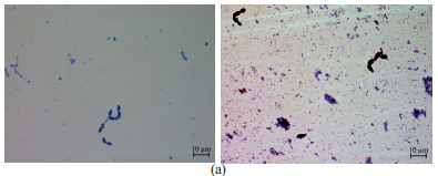
Figure 2 shows the images of biofilms formed in skimmed milk permeate on glass slides.
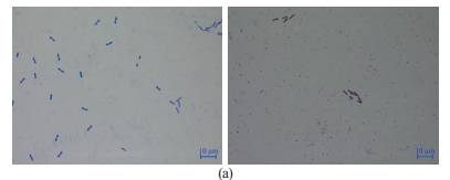
It is known that the onset of biofilm formation depends on the surface physical and chemical properties, characteristics of the initially present bacteria, and the process parameters [14, 17]. We found that biofilms in permeate samples formed rather quickly even on a smooth glass surface (Fig. 2). As early as after 4 h of the experiment, in some fields of vision, we could see individual rod-shaped and spherical cells, both purple (Gram-positive) and red (Gram-negative). However, these fields of vision were not typical and cells were not detected in a large area of glass. After 8 h, almost all fields of view featured cells of well-stained large oval (single and paired) Gram-positive cocci, as well as clusters of weakly stained small short Gram-negative rods and larger Gram-positive rods attached to the glass (Fig. 2a).
After 24 h of biofilm formation, significantly more cells remained on the glass after washing. We observed numerous clusters of spherical cells, as well as individual and long-chain sticks in separate fields of vision. Gram-stained bacteria were dark red and possibly Gram-variable. The background was a network of small bluish or pinkish rods, and their weak coloration, apparently, indicated the formation of a protective exopolymer layer (Fig. 2b). Large oval yeast cells could be seen in some vision fields, though such fields were not typical of biofilms in skimmed milk permeate.
After 48 h, a visible mucous film formed on the glass, partially washed off during the first rinse with water. Microscopic examination on glass revealed an almost continuous layer of small short Gram-negative rod-shaped bacteria with dark red cocci islands. In some fields of vision, under the upper layers of stained cells, there were dense lower layers with individual bacteria that were not visible (Fig. 2c). We can assume that the formation of a mature biofilm well fixed on the glass surface was completed at that stage.
After 72 h, we observed an increase in the mucus thickness on the glass. After rinsing with water, the micropreparations in most fields of vision looked almost the same as they did after 48 h. Large dark blue cocci, mostly paired, were visible on the surface of the biofilm. In some fields of vision, we found significant areas free of cells, which might have formed after large areas of biofilm were detached by glass rinsing (Fig. 2d). With longer incubation (up to five days), we found no significant changes in the structure of biofilms.
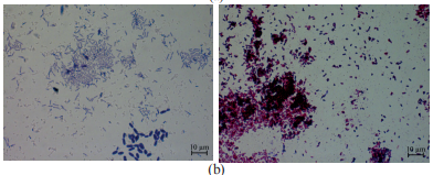
Sweet whey permeate. The samples had the following physicochemical and microbiological parameters: mass fraction of dry substances 4.9 ± 0.1%, pH 6.42 ± 0.15, NMAFAnM 1.4 ± 0.5×10³ CFU/cm3, CGB (coli-forms) 2.9 ± 0.4×10² CFU/cm3, yeast 2.8 ± 0.1×10² CFU/cm3, mold 17 ± 60 CFU/cm3.
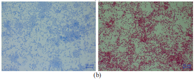
Figure 3 shows the images of biofilm formed in skimmed milk permeate on glass slides.


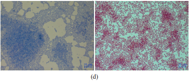
After 4 h of incubation, we found no signs of fixation or growth of microorganisms on the glasses. Microscopic examination of stained biofilms formed after 8 h showed individual cells and rare clusters of Gram-positive small cocci and Gram-negative small rods in some fields of vision (Fig. 3a). After 24 h, separate zones of Gram-positive cocci, forming pairs and short chains, appeared on a thin layer of Gramnegative short rods. We should note that during the first 24 h, biofilms in the sweet whey permeate formed more slowly than those in skimmed milk permeate (Fig. 3b). Another 24 h of incubation resulted in biofilms of approximately the same composition as in the milk samples incubated for 48 h. However, biofilms in the whey samples were less dense than those in the milk permeate (Fig. 3c).
After 72 h, the biofilm became denser, while the unevenness of its distribution increased, and spaces free of cells appeared (Fig. 3d). In many fields of vision, we found thick exopolymer layers with enclosed cells of indefinite shape. These layers were laced with long filaments of rod-shaped cells. In some areas of the biofilm, large yeast cells could be seen. Subsequently, after four and five days of biofilm formation in the sweet whey permeate, there was an increase in the mucous layer on the glass. The microstructure of biofilms after washings was similar to that formed after 72 h.
Acid whey permeate. The permeate samples had
the following physicochemical and microbiological
indicators: mass fraction of dry substances 4.8 ± 0.1%,
pH 4.65 ± 0.05, NMAOAnM 2.9 ± 0.5×10³ CFU/cm3,
CB (coli-forms) were not detected in 1 cm
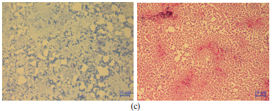
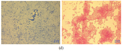
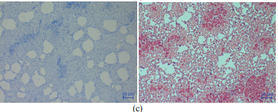
Figure 4 shows the images of biofilm formed in acid whey permeate on glass slides.


After 4 h of the experiment, we found single cells of yeast and Gram-positive cocci in some fields of vision. After 8 h, most fields of vision featured single and paired Gram-positive cocci and small clusters of large oval yeast cells (2–3×4–6 μm). We can assume that these clusters formed as a result of their longitudinal budding, sometimes with the signs of a false mycelium formation (Fig. 4a).
After 24 h, the yeast formed large groups of uniformly colored cells. Staining with methylene blue revealed than they had the clearly delineated elongated shape. We also found clusters of small rods and diplococci weakly stained with methylene blue (Fig. 4b).
After 48 h, they formed a thin broken layer with numerous clusters of yeast cells, and budding became less noticeable. In general, during this period of biofilm formation, its structure (excluding yeast cells) was similar to the structure of biofilms in skimmed milk and sweet whey permeates after 48 h. However, further incubation led to a destruction of the formed layers instead of thickening. After 72 h, we found irregularly spaced individual clusters of rod-shaped and spherical bacteria with single and grouped yeast cells (Fig. 4d). Microorganism-free zones increased. The microstructure of biofilms after four and five days was practically the same.
Some samples contained a mold resembling Geotrichum lactis in the morphology of thick hyphae, which easily disintegrated into rectangular oidiospores with rounded ends. After 48 h, a dense whitish film with a pronounced unpleasant (rancid, moldy) odor formed on the surface of the permeate in Petri dishes. This film was tightly attached to the slide and was poorly washed off.
Comparison of results of different types of raw materials and data by other researchers. The analysis of the results showed both common features and differences between the processes of biofilm formation in permeates of different types of secondary dairy raw materials on glass in statics. After 8 h, we observed the attachment of microbial cells to the glass surface and the beginning of their reproduction in all three types of raw materials. Apparently, this was the first stage of reversible attachment of microorganisms when they could be easily removed from the surface but could become the basis for further growth of biofilm [15].
After 24 h, all types of raw materials had a thin, poorly washed off biofilm formed on the glass. This process involved small Gram-negative rods, which formed a layer tightly adjacent to the glass, and Gram-positive cocci with a predominantly paired arrangement of cells located on the rod layer. Studies have shown that by this time, biofilms can already reach the stage of irreversible attachment to the surface of UF membranes, when their removal with conventional washing and disinfection protocols is difficult [16, 17].
After 48 h, a mature multilayer biofilm formed in all the permeate samples. Unevenly colored layers of biofilms and bridges between them indicated the presence of an extracellular polymeric substance. The exopolymer layer held the cells of microorganisms together, firmly attached them to the surface and protected them from adverse influences. The observed heterogeneous regions in the homogeneous matrix of the biofilm were consistent with the data obtained by Anand et al. who found that in multicultural biofilms each species could produce different polymers, which fused later [14].
At this stage, the formation of biofilm layers could occur due to the use of nutrients not only from the environment, but also from the initial film. The secretion of the exopolymer layer continued, and the thickness of the biofilm on the equipment could increase due to a continued deposition and adhesion of dairy components to the layer. This was consistent with previously published data that three-dimensional structures of mature films at a late stage of development commonly had a mushroom shape with an uneven distribution of microorganisms and channels inside. This stage of the biofilm existence might already be characterized by the processes of exchange of signaling molecules and the formation of a “quorum sensing”, the exchange of genetic information and the acquisition of inherited resistance to antibiotics and disinfectants [4, 14].
Later (after 72 h or more), rinsing the glasses separated part of the mucus formed on their surface. All the samples showed areas free from microorganisms. Apparently, this occurred due to partial destruction of a biofilm, which corresponded to the last stage of their development [4, 14].
The main difference between biofilms in sweet and acid whey was their slower formation at the first stage (24 h) compared to biofilms in skimmed milk. This might be due to its composition (for example, the higher protein content in milk) and the properties of the raw materials (for example, the lower active acidity of whey compared to milk). Perhaps these differences were due to the influence of starter cultures contained in these types of raw materials, which were based on Lactococcus lactis subsp. lactis capable of producing nisin.
The obtained data were consistent with the findings of Marka and Adand [15]. The authors found that during the processing of skimmed milk permeate, biofilms on reverse osmosis membranes formed faster than during processing of sweet whey. Apparently, this was due to the fact that the microflora of milk is represented by α-, β- and γ-proteobacteria, bacilli, flavobacteria and actinobacteria, while fresh and pasteurized sweet whey mainly contains Lactococcus spp. [16]. The structural peculiarity of biofilms in the acid whey permeate was yeast, which took part in their formation at all stages of their development. Probably, due to autolysis and the release of various enzymes into the environment, yeast contributed to the destruction of biofilms after 72 h. We should note that the role of yeast in biofilm formation has been little studied and requires further analysis.
In the later stages of development, the upper layers of biofilms became unstable and were easily detached when the glasses were rinsed. This can cause continuous contamination of raw materials and products. At the same time, the cells separated from a biofilm are more resistant to external influences as compared to ordinary (planktonic) cells.
Studies have shown that the formation of biofilms can lead to biodegradation of materials, equipment malfunctions, a decrease in its performance, increased energy consumption and consumption of detergents, as well as problems with the quality and safety of products [2, 4, 14]. The peculiarities of the composition and properties of biofilms in different types of raw materials must be taken into account when improving the procedures of washing technological equipment with alkalis, acids and enzymes.
Removal of biofilms formed in secondary dairy raw materials on glass. To study the patterns of biofilm removal, we used the standard procedure of washing the electrodialysis unit and the spectrophotometric method for determining the optical density of the washings. When setting up the experiments, we assumed that the upper layers of the biofilm, which were not attached to the glass surface, passed into a wash water after the first rinsing of the glasses. The fixed layers of biofilms were removed with cleaning solutions (acidic, alkaline and enzymatic). After using each type of washing solution, the slides were washed with distilled water.
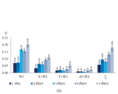
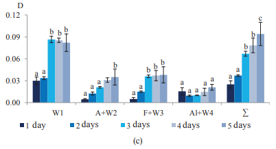
The washout results are presented in diagrams (Fig. 5). They show the optical density (D) of the wash water after the first rinsing of the glasses (W1), the total D of the acid solution and the subsequent wash water (A + W2) after rinsing glasses; the total D of the enzyme solution and the subsequent wash water (F + W3) after rinsing glasses; the total D of the alkali solution and the subsequent rinsing water (Al + W4) after rinsing the glasses, as well as the total D (Σ) for all washings of the fixed layers of biofilm with washing solutions (A + W2, F + W3 and Al + W4).
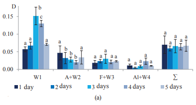
Skimmed milk permeate. The analysis of results (Fig. 5a) shows that the amount of structural elements of biofilms formed on glass in the permeate, easily washed off with water, increased significantly after three and four days, and by five days it decreased to the level of the first and second days.
The acidic wash solution was more effective in removing microorganisms from the 24 h biofilm than on the following three days. The efficiency of enzyme solutions was somewhat lower, and the lowest values of optical density were recorded in alkaline solutions. The maximum values of the optical density of washings in enzyme solutions were found after three days, in alkaline solutions – after four days. However, we did not reveal a statistical significance of differences in the optical density of washings in these solutions on different days of the experiments.
The total optical density of washing solutions remained practically constant for five days of the experiment. This indicated that as early as after 24 h, a layer of well-fixed microorganisms with a fairly stable structure and thickness formed on the glass in the skimmed milk permeate. The properties of this layer changed little in the following days, while layers of microorganisms gradually built up on top, which were easily washed off with water. The thickness of these layers reached its maximum after three days.
A microscopic examination of glass slides after all stages of washing in some fields of vision revealed individual cells of cocci and rods.
Sweet whey permeate. We found that in the first two days, the average values of the optical density of the washings after the first rinsing of glasses with water were similar to those obtained in experiments with skimmed milk, but with a wider range of values (Fig. 5b). However, after three and five days of experiments, much more microorganisms were washed off from biofilms in sweet whey permeate than from biofilms in milk permeate. A significant decrease in the optical density values of these washings after four days indicated the instability of the properties of the upper layers of mature biofilms.
Acid treatment was more effective in removing sweet whey biofilms than other detergent solutions. At the same time, there was a gradual increase in the values of optical density of washings in acid solution from 1 to five days, with a slight decrease after three days. Apparently, this was due to the growth of fixed exopolymer layers of biofilms, which were soluble in an acidic medium. The optical density of the washings in enzyme and alkaline solutions was lower than in acidic solutions, and remained at the same level throughout the experiment, except a statistically significant increase in the enzyme solution after five days. Assumingly, few microorganisms remained on the glass after acid washing. However, microscopic examination of the slides after all stages of washing revealed a significant number of microorganisms, including cocci, yeast, and in some fields of view, clusters of rods.
The analysis of the total values of the optical density of washing solutions showed that the fixed layer of biofilms increased significantly after two, four and five days. The 3-day biofilm was more unstable, with part of it removed during the first washing. In general, fixed and non-fixed layers of biofilms in sweet whey permeate tended to grow during the entire experiment, reaching their maximum after five days.
Acid whey permeate. The experiments with acid whey revealed lower values of the optical density of washings after the first rinsing of glasses than in the experiments with other types of secondary dairy raw materials (Fig. 5c). Despite a significant increase in optical density after three and five days, it was approximately 1.5 and 2 times lower than in skimmed milk and in sweet whey, respectively.
The cleaning efficiency with acidic and enzyme solutions gradually increased with the growth of the biofilm. We found a statistically significant increase in the optical density of the washings on day five for acidic solutions and on days three and five for enzyme solutions. Alkaline treatment of a 24 h biofilm was more effective than in acidic and enzyme solutions. This suggested that the properties of biofilms in acid whey at the initial stage of development differed from those of biofilms in skimmed milk and sweet whey. Subsequently, with an increase of biofilm formation time, the efficiency of acidic and enzymatic treatments increased.
The total optical density of washings from biofilms in the acid whey permeate on the first day was 3 and 2.4 times lower than in similar experiments with skimmed milk and sweet whey, respectively. After five days of biofilm formation in the acid whey permeate, the total optical density reached its maximum values, which was 1.3 times higher than in milk permeate and two times lower than in sweet whey permeate.
Microscopic examination of glass slides after all cleaning procedures revealed cells of microorganisms, including yeast, and in some experiments, milk mold.
Comparison of results of different raw material types and data by other researchers. We found that at the first stages of biofilm formation, all studied types of secondary dairy raw materials showed similar general patterns of biofilm removal during the first washing with water. All the species demonstrated the same level of optical density after one and two days, with its significant increase after three days. At the same time, the highest washout values during research period were found in experiments with sweet whey, the lowest one in experiments with acid whey.
The remaining layers of biofilms were effectively removed in acid solutions, which was characteristic of the experiments with sweet whey permeate. These results were consistent with the data of other researchers who studied the processes of washing ultrafiltration and reverse osmosis membranes used for processing secondary dairy raw materials [14].
Alkaline solutions are known to be widely used in the dairy industry to dissolve and hydrolyze organic substances [4]. In our work, alkaline solutions were ineffective in removing biofilms for all studied samples. Perhaps it was due to the fact that by this time of processing, fewer structural elements of biofilms remained on the glass. Moreover, cells of microorganisms were found on the glasses in all experiments, after all procedures of removing biofilms. Their number gradually increased even when the treated glasses were stored in sterile distilled water. This indicated their viability and a possibility of a new biofilm to form.
Interestingly, fixed biofilm layers formed in a skimmed milk permeate as early as after 24 h, and their thickness and properties hardly changed over the next four days. In contrast to this, fixed biofilm layers in the permeates of sweet and acid whey gradually increased during the entire time of biofilm formation.
The fastest and slowest growth of biofilms, especially after three to five days, was found in sweet whey and in acid whey, respectively. Microorganisms of whey permeates formed the most firmly fixed biofilms, for the removal of which the washing procedures were insufficient. According to Marka and Anand, biofilms of sweet whey were less resistant to standard CIP washing than biofilms of skimmed milk. Noteworthily, these results were obtained in the study on reverse osmosis membranes, with the processing of raw materials lasting 48 hours and the washing conditions being different [15].
We should also note that the conditions of modeling biofilms removal from glass used in this work did not fully correspond to real production conditions. The efficiency of cleaning production equipment does not only depend on the type, dosage and pH of the detergent, but also on pressure, washing time, flow rate during washing, and solution temperature [14]. In this regard, it would be interesting to research biofilms formed in different types of raw materials under industrial conditions.
ВЫВОДЫ
The study revealed general regularities and differences in the formation and removal of biofilms formed on glass in statics in three types of secondary dairy raw materials – skimmed milk permeates, sweet whey and acid whey permeates.
The main general regularities included:
– time needed for primary attachment of
microorganisms to glass (about 8 h), the formation of
mature unevenly colored multicultural biofilms (about
48 h), and their partial destruction (72 h and more);
– bacteria participating in the formation of biofilm
structure, similar in morphological and tinctorial
properties, Gram-negative rods and Gram-positive cocci
with a predominantly paired arrangement of cells;
– peculiarities of biofilm removal at the first stages of
their formation during the first washing of glasses with
water; and
– viable cells of microorganisms detected on glasses
after all biofilm removal procedures.
The main differences were as follows:
– biofilms formed slower in the permeates of sweet and
acid whey at the first stage (24 h) compared to biofilms
in skimmed milk permeate;
– yeast participated in the formation of biofilms in the
acid whey permeate at all stages of their development
compared to the other permeates;
– the fixed layers of biofilms had higher stability in
skimmed milk compared to those in whey;
– biofilms in sweet whey demonstrated a more
significant growth after 35 days than those in skimmed
milk and acid whey.
The revealed features of biofilm composition and properties in different types of secondary dairy raw materials should be taken into account to improve the procedures of washing technological equipment used in their processing.
КОНФЛИКТ ИНТЕРЕСОВ
The authors declare that there is no conflict of interest related to this article.БЛАГОДАРНОСТИ
The authors are grateful for assistance in microscopic examination of preparations to the Research and Training Laboratory for Experimental Immunomorphology, Immunopathology and Immunobiotechnology of the Institute of Life Science, North Caucasus Federal UniversityФИНАНСИРОВАНИЕ
The work was carried out within the framework of the grant from the Ministry of Science and Higher Education of the Russian Federation (Minobrnauka) (contract № 03.G25.31.0241).СПИСОК ЛИТЕРАТУРЫ
- Flemming H-C, Wingender J, Szewzyk U, Steinberg P, Rice SA, Kjelleberg S. Biofilms: an emergent form of bacterial life. Nature Reviews Microbiology. 2016;14(9):563–575. https://doi.org/10.1038/nrmicro.2016.94.
- Galié S, García-Gutiérrez C, Miguélez EM, Villar CJ, Lombó F. Biofilms in the food industry: health aspects and control methods. Frontiers in Microbiology. 2018;9. https://doi.org/10.3389/fmicb.2018.00898.
- Coughlan LM, Cotter PD, Hill C, Alvarez-Ordóñez A. New weapons to fight old enemies: novel strategies for the (bio)control of bacterial biofilms in the food industry. Frontiers in Microbiology. 2016;7. https://doi.org/10.3389/fmicb.2016.01641.
- Hoong Teh K, Flint S, Brooks J, Knight G. Biofilms in the dairy industry. Wiley Blackwell; 2015. 288 p.
- Ryabtseva SA, Hramtsov AG, Evdokimov IA, Anisimov GS, Salova OV. Biofilms in the dairy industry: significance, formation, control. Dairy Industry. 2018;(1):57–59. (In Russ.). https://doi.org/10.31515/1019-8946-2018-1-57-59 .
- Mardanova AM, Kabanov DA, Rudakova NL, Sharipova MR. Bioplenki: osnovnye printsipy organizatsii i metody issledovaniya [Biofilms: basic principles of organization and research methods]. Kazan: Kazan Federal University; 2016. 42 p. (In Russ.).
- Rossi C, Chaves-López C, Serio A, Goffredo E, Goga BC, Paparella A. Influence of incubation conditions on biofilm formation by Pseudomonas fluorescens isolated from dairy products and dairy manufacturing plants. Italian Journal of Food Safety. 2016;5(3):154–157. https://doi.org/10.4081/ijfs.2016.5793.
- Quintieri L, Fanelli F, Caputo L. Antibiotic resistant Pseudomonas spp. Spoilers in fresh dairy products: an underestimated risk and the control strategies. Foods. 2019;8(9). https://doi.org/10.3390/foods8090372.
- Ostrov I, Sela N, Belausov E, Steinberg D, Shemesh M. Adaptation of Bacillus species to dairy associated environment facilitates their biofilm forming ability. Food Microbiology. 2019;82:316–324. https://doi.org/10.1016/j.fm.2019.02.015.
- Bassi D, Cappa F, Gazzola S, Orrù L, Cocconcelli PS. Biofilm formation on stainless steel by Streptococcus thermophilus UC8547 in milk environments is mediated by the proteinase PrtS. Applied and Environmental Microbiology. 2017;83(8). https://doi.org/10.1128/AEM.02840-16.
- Salas-Jara MJ, Ilabaca A, Vega M, García A. Biofilm forming Lactobacillus: new challenges for the development of probiotics. Microorganisms. 2016;4(3). https://doi.org/10.3390/microorganisms4030035.
- Thiran E, Di Ciccio PA, Graber HU, Zanardi E, Ianieri A, Hummerjohann J. Biofilm formation of Staphylococcus aureus dairy isolates representing different genotypes. Journal of Dairy Science. 2018;101(2):1000–1012. https://doi.org/10.3168/jds.2017-13696.
- Marti R, Schmid M, Kulli S, Schneeberger K, Naskova J, Knøchel S, et al. Biofilm formation potential of heat-resistant Escherichia coli dairy isolates and the complete genome of multidrug-resistant, heat-resistant strain FAM21845. Applied and Environmental Microbiology. 2017;83(15). https://doi.org/10.1128/AEM.00628-17.
- Anand S, Singh D, Avadhanula M, Marka S. Development and control of bacterial biofilms on dairy processing membranes. Comprehensive Reviews in Food Science and Food Safety. 2014;13(1):18–33. https://doi.org/10.1111/1541-4337.12048.
- Marka S, Anand S. Feed substrates influence biofilm formation on reverse osmosis membranes and their cleaning efficiency. Journal of Dairy Science. 2017;101(1):84–95. https://doi.org/10.3168/jds.2017-13249.
- Chamberland J, Lessard M-H, Doyen A, Labrie S, Pouliot Y. A sequencing approach targeting the 16S rRNA gene unravels the biofilm composition of spiral – wound membranes used in the dairy industry. Dairy Science and Technology. 2016;96(6):827–843. https://doi.org/10.1007/s13594-016-0305-2.
- Chamberland J, Lessard M-H, Doyen A, Labrie S, Pouliot Y. Biofouling of ultrafiltration membranes by dairy fluids: Characterization of pioneer colonizer bacteria using a DNA metabarcoding approach. Journal of Dairy Science. 2017;100(2):981–990. https://doi.org/10.3168/jds.2016-11829.
- Ryabtseva SA, Tabakova YuA, Anisimov GS, Kravtsov VA, Salova OV. Formation and removal of biofilms in skimmed milk permeate. Dairy Industry. 2019;(6):48–50. (In Russ.). https://doi.org/10.31515/1019-8946-2019-6-48-50.
