

Abstract
The research featured the effect of a diet fortified with essential microelements on the ruminal microbiota of young rams. Ruminal microbiota is largely responsible for feed digestibility and body functioning of cattle.The study involved the contents of the rumens and jejuna of seven-month-old rams of the Edilbaev breed, which were subjected to a biofortified diet. The diet included the Russian feed additives Yoddar-Zn and DAFS-25 represent a protein-carbohydrate complex with plant silicon. The microflora of the digestive tract was tested using the molecular genetic method of terminal restriction fragment length polymorphism (T-RFLP) sequestration. The microstructural studies of the jejunum samples exploited light microscopy.
The feed additives increased the population of cellulolytic and lactate-fermenting bacteria, as well as the Prevotella sp. microbiome and bifidobacteria in the rumen samples. The data obtained revealed the effect of essential microelements on the taxonomic pattern of microorganisms and the microflora profile. The research revealed the ratio of normal, opportunistic, pathogenic, nonculturable, and transit microflora. The jejunum wall samples obtained from the experimental group that fed on Yoddar-Zn and DAFS-25 had a more distinct micropicture of mucous membrane. Their rumen microflora balance had fewer pathogenic and opportunistic microorganisms, which was also confirmed by the jejunum morphology.
The feed additives DAFS-25 and Yoddar-Zn proved beneficial for ram diet and inhibited the negative effect of pathogenic treponemas on the rumen. The additives improved digestion, absorption, and assimilation of food nutrients, as well as increased the livestock yield.
Keywords
Young rams, animal diet, feed additives, essential microelements, molecular genetics, jejunum, microbiocenosis, microstructural studiesINTRODUCTION
Feed composition has a direct impact on the qualitative and quantitative characteristics of the gastrointestinal microbial community. Minerals and vitamins are essential micronutrients that participate in such vital processes as enzyme formation or the synthesis and metabolism of hormones and vitamins. They affect the nervous, cardiovascular, and endocrine systems, as well as the activity of the endocrine glands and the gastrointestinal tract.
Micronutrient deficiency may trigger various infectious and non-infectious diseases [1, 2]. A poorlybalanced feed ration often leads to undesirable changes in the microbiota of small ruminants. The resulting digestive disorders cause various diseases and eventually lead to poor livestock yield. Biofortification fortifies animal diet with essential nutrients, thus improving the chemical composition of meat. It renders high-quality mutton that provides consumers with essential microelements [3–9].
Practical microbiology gives scientific data on the composition, role, or function of the microbial community in the rumen content of small ruminants. However, some of these methods have disadvantages or limitations. For instance, researchers cannot choose the optimal environment for microbial cultivation. Fortunately, contemporary molecular genetic methods make it possible to skip the stage of cultivation and study microorganisms without the restrictions that traditional diagnostic microbiology are prone to [10–14].
Small intestine (lat. intestinum tenue) of farm animals absorbs nutrients from the chyme. It is in the small intestine that the main digestion takes place, and this is where most digestive enzymes come from. Partially digested food leaves the stomach and enters the duodenum, where it is processed by intestinal and pancreatic juices and bile. The small intestine is where digested food, toxins, poisons, medicinal substances, etc. are absorbed into the bloodstream or lymphatic channel [15–19].
The jejunum is somewhat structurally different from other parts of the small intestine. Membrane digestion is at its utmost in the upper parts of the jejunum. As a result, its wall is thicker; it has more folds in the mucous membrane, denser villi, and a more abundant blood supply [20–22]. Therefore, the small intestine is a vital system of animal body, and its flawless work is essential for sheep farming, which proves the relevance of this research.
Sheep farming needs new fundamental data on the effect of biofortification on the bacterial rumen community. Bacterial profile includes normal, opportunistic, and pathogenic microflora, as well as nonculturable and transit microflora that does not affect the life of the animal. Light microscopy revealed the morphology of the intestine and the main differences between the samples obtained from animals fed with Yoddar-Zn and DAFS-25.
The research objective was to assess the effect of essential microelements on the ruminal microbiocenosis and the microstructure of the jejunum in young rams.
STUDY OBJECTS AND METHODS
The next-generation sequencing (NGS) revealed the digestive microflora of seven-month-old rams of the Edilbaev breed. The experiment made it possible to evaluate the effect of the feed additives Yoddar-Zn (Material Specifications TU 10.91.10-252-10514645- 2019) and DAFS-25 (Material Specifications TU 10.91. 10-253-10514645-2019). The studies took place in the laboratory of molecular genetic research of the Research and Production Company BIOTROF (St. Petersburg, Russia).
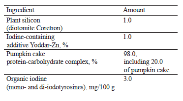
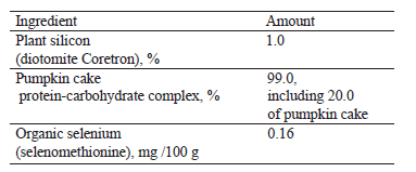
The feed additives were developed at the Volga Region Research Institute for the Production and Processing of Meat and Dairy Products. Both feed additives contain Coretron, an enterosorbent used in Russia in cattle diet, and cold-pressed pumpkin cake, which served as a protein-carbohydrate component (Tables 1 and 2) [6].
A scientific and economic experiment was necessary to assess the effectiveness of various diets fortified with organic microelements, i.e., mono- and di-iodotyrosines and selenomethionine. After weaning from mothers at the age of four months, 100 lambs of the Edilbaev breed were divided into four groups, 25 animals in each. The lambs were fed and fattened in the same way. On day 105, when the animals were seven months old, they were slaughtered by the traditional method according to the Technical Regulations of the Customs Union on the safety of meat and meat products TR TS 034/2013. Prior to slaughter, all experimental animals had received no food for 24 h.
Yoddar-Zn is a source of bioavailable organic iodine and zinc. It also contains iodized milk proteins associated with amino acids and zinc compounds. Yoddar-Zn owes its biological properties to bound iodine, which is necessary for the biosynthesis of such thyroid hormones as thyrotoxin and triiodothyropine. They are important for metabolism and immune system [6].
The control group of young rams received 300 grams of mixed fodder per head per day. The first experimental group received daily the same mixed fodder together with 300 mg of Yoddar-Zn, the second experimental group – 0.5 mg of DAFS-25, and the third experimental group – a mix of these additives (300 and 0.5 mg).
The effect of the organic additives was studied in vivo by comparing the microbiocenosis and microstructural parameters of the small intestine in the experimental and control groups of young rams.
The next generation sequencing (NGS) is currently one of the most optimal research methods. NGS technologies provide metagenomic studies of complex microbial communities with a large volume of read nucleotide sequences. This technology is much more accurate than the Sanger sequencing in determining the phylogenetic species of microorganisms [23].
The in vivo assessment of the impact on the intestinal microbiocenosis took 105 days. Samples of the rumen contents were put into sterile containers (Pan Eco, Russia) immediately after the slaughter and tested for microbial composition. Next step included histology of jejunum samples. The preparations were stained with hematoxylin and eosin to assess any possible changes in the intestinal mucosa.
The bacterial content of the ram rumen was analyzed by NGS method. Total DNA was isolated by using the Genomic DNA Purification Kit (Fermentas, Inc., Lithuania) according to the manual. The final concentration of total DNA in the solution was measured using a Qubit fluorimeter (Invitrogen, Inc., USA) with Qubit dsDNA BR Assay Kits (Invitrogen, Inc., USA) according to the manual.
The NGS was performed on a second-generation MiSeq sequencing platform (Illumina, Inc., USA) with primers for the V3-V4 region of 16S rRNA; upstream primer – 5´-TCGTCGGCAGCGTCAGATGTGTATAAG AGACAGCCTACGGGNGGCWGCAG-3´; downstream primer – 5´-GTCTCGTGGGCTCGGAGATGTGTATA AGAGACAGGACTA-CHVGGGTATCTAATCC-3´ [24].
Libraries were prepared with Nextera® XT IndexKit reagents (Illumina, Inc., USA); the PCR products were purified with Agencourt AMPure XP (Illumina Inc., USA); the sequencing was performed with MiSeq® ReagentKit v2 (500 cycle) (Illumina, Inc., USA) [25].
The obtained reads underwent overlapping, filtering by Q30 quality, and primer trimming. The processing involved the Illumina bioinformatics platform (Illumina, Inc., USA). The quality control and assessment of the taxonomic composition were carried out using the QIIME2 v.2019.10 software (https://docs.qiime2.org) and the Green-Genes database 13.5 (https://greengenes.secondgenome.com).
Pieces of ram jejunum samples were removed by preparation and fixed in 10% aqueous neutral formalin solution at room temperature for 48 h. The selected samples were removed from the fixing liquid and washed under running water for 48 h. For dehydration, the material was washed in alcohols of increasing concentration from 50 to 96%. After that, the material was embedded in paraffin shaped in paraffin blocks. Sections of 5–8 μm were sliced with a sledge microtome, deparaffinized, and stained by Ehrlich hematoxylin and eosin dyes. Hematoxylin stains basophilic cellular elements bright blue, while eosin alcohol acid dye stains Y-eosinophilic cell elements pink. Basophilic structures most often contain nucleic acids (DNA and RNA), i.e., nucleus, ribosomes, and RNA-containing cytoplasm sections. Eosinophilic elements contain intra- and extracellular proteins. Cytoplasm belongs to the main eosinophilic environment, so its elements stain bright red [1].
Microscopy involved a Levenhuk MED PRO 600 Fluo microscope, which is designed for transmitted light brightfield microscopy or for a luminescent (fluorescent) method (Magnification ×300).
The morphometric analysis of the obtained data traced the thickness of the jejunum layers. The experiment relied on a screw eyepiece micrometer MOV-1-15× and an eyepiece ruler with 60 units of scale division. The quantitative parameters of the histological structures underwent further statistical processing.
Statistical processing of the obtained digital data followed standard methods using the Microsoft Excel 2010 (Microsoft Corp., USA) and the statistical data analysis package StatPlus 2009 Professional 5.8.4 for Windows (StatSoft, Inc., USA). Student’s t-test was applied to assess the reliability of data between the experimental and control groups.
RESULTS AND DISCUSSION
This section describes the effect of feed additives Yoddar-Zn and DAFS-25 in the diet of young Edilbaev breed rams on their ruminal microbiocenosis and jejunum microstructure.
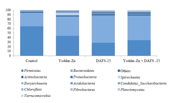
The NGS analysis revealed the ruminal bacteria community in the control and experimental groups. The rumen samples contained 31 phyla of bacteria and 1 phylum of archaea (Fig. 1). Firmicutes and Bacteroides predominated with a total share of 86–94%. The share of Actinobacteria, Spirochaetes, and Candidatus Saccharibacteria was 1–6%. In the control group, Firmicutes ranked first: their relative value in the community was 65%, while the proportion of Bacteroides was only 29.4%. This ratio was different in the experimental groups. In the group that received Yoddar-Zn, the proportion of Firmicutes and Bacteroides was the same (42–43%). In the groups that received DAFS-25 and DAFS-25 + Yoddar-Zn, the ratio of these two phyla was reversed compared to the control group: Bacteroides – 50–60%, Firmicutes – 30–35%.
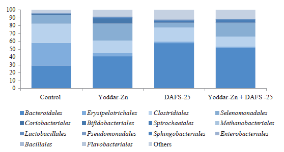
At the level of orders, the community was dominated by Bacteroidales, Erysipelotrichales, and Clostridiales. Rams fed with DAFS-25 had a larger proportion of Bifidobacteriales (5.8%). The control group had more Erysipelotrichales – 28.8%.
Cellulolytic bacteria are important bacterial community members. They break down the fiber of plant foods and convert it to volatile fatty acids. Cellulolytic bacteria in the rumen samples were mainly represented by the bacterial families Clostridiaceae, Prevotellaceae, Flavobacteriaceae, Eubacteriaceae, Lachnospiraceae, Ruminococcaceae, and Thermoanaerobacteraceae, as well as by the Bacteroidetes phylum.
The total proportion of cellulolytic bacteria was different in all samples. The share of beneficial cellulolytic bacteria ranged from 51.3 to 75.4%, depending on the sample. The control group had the smallest proportion of cellulolytic bacteria, while the group that received DAFS-25 had the largest one. In the groups treated with Yoddar-Zn and DAFS-25 + Yoddar- Zn, the proportion of cellulolytic bacteria was 56.6 and 64.1%, respectively.
Lactate-utilizing bacteria are another important group in the ruminal bacterial community. They ferment lactic acid produced by bacteroids and lactic acid bacteria and other organic acids into volatile fatty acids used in metabolic processes.
The NGS analysis showed that the content of Veillonellaceae lactate-utilizing bacteria was very large in some samples. In the groups that received Yoddar- Zn and DAFS-25 + Yoddar-Zn, their content was 20.6 and 12.9%, respectively, while the control group and the experimental group fed with DAFS-25 alone, it was 9.1 and 5.1%, respectively. This indicator may demonstrate that these bacteria are especially active in the sheep rumen, depending on their physiological state of the animal.
The share of bacterial pathogens was insignificant in all samples and totaled about 0.5% in all groups. Opportunistic Enterobacteriaceae were also represented in a very small amount (≤ 0.1%) in all samples.
Prevotella appeared to be the dominant genus. Its relative abundance in the experimental groups exceeded the control (28.3, 38.9, and 33.4 vs. 22.8%). Prevotella sp. often is the most numerous genera in sheep rumen. For instance, Prevotella also dominated in a similar study by Cui et al. on the effect of selenium feed additives on the microbial community in sheep [3]. Cui et al. also proved the significant effect of selenium on ruminal bacterial populations and microbial fermentation in the rumen in general.
Subdominant microorganisms in the rumen were represented by the Dysgonomonas, Saccharofermentans, Tangfeifania, and Treponema genera. Cui et al. showed that the abundance of Saccharofermentans sp. was in inverse relationship with selenium. Our research, on the contrary, proved that the amount of Dysgonomonas sp. and Prevotella sp. depended on the presence of selenium in the diet.
To identify and evaluate the changes in the small intestine wall, jejunum wall pieces were subjected to microscopy [1].
This research of the effect of biofortification on the microstructure of sheep jejunum yielded a more accurate assessment of the safety of Yoddar-Zn and DAFS-25 for small rumens [7, 8].
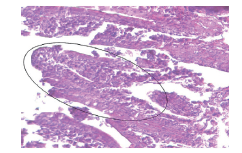
Light microscopy of the jejunum in all samples revealed that the mucous membrane was well-structured, with distinct layers. The mucous membrane of the jejunum consisted of four layers: innermost mucosa outermost, submucosa, muscularis (outer and inner layers), and serosa. The columnar villi (Fig. 3) of the mucosal epithelial layer were distinct and consisted of a single-layer columnar epithelium lining the crypts. The structure of the layer was dominated by goblet cells and limbic epithelial cells, which produce mucus. The lamina propria consisted mostly of cells and fibers of loose fibrous connective tissue. The muscular layer was represented by two distinct alternating layers of myocytes: annular and longitudinal. The submucosa was represented by loose fibrous tissue with clear contoured blood and lymphatic vessels, as well as complex tubularalveolar glands that produced intestinal juice.
The muscular membrane of the jejunum tissue had two distinct layers of myocytes, which were separated by a minimal layer of connective tissue. The structure was clear; the cells were elongated and spindle-shaped.
On the outside, the jejunum was covered with a serous membrane with layers of loose connective tissue and mesothelium. The integrity of the latter was intact.
Figure 3 shows the mucous membrane of the jejunum samples in the control group. The general histological structure remained the same. We observed a slight accumulation of mucus between the villi produced by goblet cells. Epithelial cells were of an elongated cylindrical shape. The glands of the lamina propria were well expressed. The integrity of the layers was intact.
The jejunum samples in the experimental groups had some histological features that differed from the control group samples.
The jejunum of young rams that received Yoddar-Zn had a single-layer cylindrical border epithelium on the transverse sections of the villi (Fig. 4).
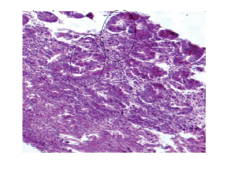
The lumen of the tubular glands looked deserted, and the crypts were separated by a minimal layer of connective tissue (Fig. 4). The muscular plate of the mucosa was well expressed; the submucosa consisted of connective tissue layers with elongated tubular glands. The integrity of all membranes was intact.
Figure 5 shows the jejunum samples obtained from animals that received DAFS-25. The cylindrical epitheliocytes and the villi of the lamina propria were distinct, with moderately pronounced glands with empty lumens and numerous goblet cells. The integrity of the membranes was intact: the muscle layers were separated from each other by connective tissue. The serous tissue was hardly developed.
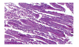
Figure 6 shows the jejunum samples obtained from animals that received DAFS-25 + Yoddar-Zn. The organ wall had a very obvious microstructure. The structure of the mucous membrane of the small intestine was intact, its constituent elements having clear contours. The goblet cells and the single-layered columnar epithelium were quite distinct. The villi were separated from each other by a minimal layer of connective tissue. The submucosa demonstrated contoured blood vessels, some of which were filled with blood. This fact indicates a more intensive metabolism in animals fed with DAFS- 25 + Yoddar-Zn.
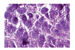
The myocytes of the muscular membrane are quite clearly separated by loose fibrous connective tissue with a minimal number of blood vessels. Muscle cells corresponded to the state of contraction, i.e., the cells were as if the muscle was contracted, and the morphology of the early autolysis process.
The morphological analysis proved that the structure of the jejunum wall in the control and experimental groups was intact and typical. The layers had an integral structure in all experimental groups. Samples obtained from animals that received DAFS-25 + Yoddar-Zn had the best developed structure.
Table 3 shows that the arithmetic mean value of the thickness of the jejunum mucous layer was 19.40 ± 0.55 μm in the rams of the experimental groups, which exceeded the control by 2.0 μm. The thickness of the muscular membrane in experimental groups also exceeded this indicator in the control group by an average of 0.8–2.0 μm. The experimental rams also had a slightly thicker serous layer.
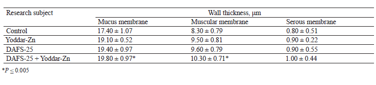
The minimal thickening of the jejunum membranes was minimal in the experimental groups, the lowest observed in the animals that received DAFS-25 + Yoddar-Zn. This fact may be an indirect indicator of a more active digestion, a better digestibility, and a greater absorption of feeds and nutrients into the bloodstream.
CONCLUSION
Biofortification of young rams’ diet with essential microelements had a positive effect on the quality and quantity of the gastrointestinal microbial community, which means a better digestion process and a greater animal yield.
In the rumen samples, cellulosolytic bacteria, which break down the fiber of plant foods into volatile fatty acids, were mainly represented by Clostridiaceae, Prevotellaceae, Flavobacteriaceae, Eubacteriaceae, Lachnospiraceae, Ruminococcaceae, and Thermoanaerobacteraceae families, as well as by the Bacteroidetes phylum. The content of lactate-utilizing bacteria in the rumen samples reached 40%, which may indicate a high degree of activity of these bacteria, depending on their physiological state of the animal.
The content of bacilli in the rumen samples was ≤ 1%. The total proportion of pathogenic species ranged from 0.2 to 6.3%. The experiment revealed ≥ 50 types of pathogenic microorganisms, which were most abundant in the group fed with Yoddar-Zn + DAFS-25. The pathogenic microorganisms belonged to erysipelothrix, fusobacterial, and streptococci. The content of porphyromonas reached 0.68% of total microorganisms, while the proportion of Treponema in the samples ranged from 0.6 to 1%. Lactobacilli were represented mainly by Lactobacilliales (0.06–0.45%). This fact may indicate a high degree of activity of these bacteria in the sheep rumen, depending on their physiological state of the animal.
The balance of the microflora in the sheep rumen samples was good, and the amount of beneficial microflora was enough to inhibit the pathogenic and opportunistic bacteria.
The light microscopy revealed no adverse effect of the feed additives DAFS-25 and Yoddar Zn on the microstructural parameters of sheep jejunum. Therefore, they can be recommended for fattening purposes in industrial conditions.
The additives had no negative impact on the rumen microbiocenosis and the jejunum microstructure. The structure of the jejunum corresponded to the morphological characteristics for this type and age of farm animal in all the groups. A clearer micropicture of the jejunum wall was revealed in the experimental group of rams fed with DAFS-25 + Yoddar Zn.
The complex application of additives DAFS-25 and Yoddar Zn helped optimize the processes of digestion, absorption, and assimilation of feed nutrients, which was partly confirmed by the minimal thickening of the jejunum membranes.
Further research is needed to study the effect of these additives on other important systems of animal organism, e.g., digestive (liver), excretory (kidneys), nervous (cortex and base of brain), and immune (spleen and mesenteric lymph nodes) systems.
Contribution
Authors are equally relevant to the writing of the manuscript, and equally responsible for plagiarism.CONFLICTS OF INTEREST
The authors declare no conflict of interest.FUNDING
The research was financed by the Russian Science Foundation, Project No. 19-76-10013 “Development and implementation of production and storage technology for environmentally friendly mutton fortified with essential microelements”.REFERENCES
- Khvylya SI, Giro TM. Assessment of the quality and biological safety of meat and meat products by microstructural methods. Saratov: Bukva; 2015. 240 p. (In Russ.).
- Ozer N, Birişik C, Sakata R, Yetim H, Ahhmed MA. Meat therapy for hypertension: hybrid hydrolysate as ace inhibitory compounds. Proceeding of the 61st international congress of Meat Science and Technology; 2015. Clermont. Clermont; 2015. p. 108–111.
- Cui X, Wang Z, Tan Y, Chang S, Zheng H, Wang H, et al. Selenium yeast dietary supplement affects rumen bacterial population dynamics and fermentation parameters of Tibetan sheep (Ovis aries) in alpine meadow. Frontiers in Microbiology. 2021;12. https://doi.org/10.3389/fmicb.2021.663945
- Kulikovskii AV, Lisitsyn AB, Chernukha IM, Gorlov IF, Savchuk SA. Determination of iodotyrosines in food. Journal of Analytical Chemistry. 2016;71(12):1215–1219. https://doi.org/10.1134/S1061934816100087
- Bo Trabi E, Seddik H, Xie F, Wang X, Liu J, Mao S. Effect of pelleted high-grain total mixed ration on rumen morphology, epithelium-associated microbiota and gene expression of proinflammatory cytokines and tight junction proteins in Hu sheep. Animal Feed Science and Technology. 2020;263. https://doi.org/10.1016/j.anifeedsci.2020.114453
- Giro TM, Kulikovsky AV, Knyazeva AS, Domnitsky IYu, Giro AV. Biochemical and microstructural profile of the thyroid gland from lambs raised on experimental diets. Food Processing: Techniques and Technology. 2020;50(4):670–680. (In Russ.). https://doi.org/10.21603/2074-9414-2020-4-670-680
- Giro TM, Kulikovski AV, Giro VV, Mosolov AA. Microstructural studies of muscle tissue of lamb of aboriginal breeds of the Volga region. IOP Conference Series: Earth and Environmental Science. 2020;548(8). https://doi.org/10.1088/1755-1315/548/8/082082
- Chernukha IM, Mashentseva NG, Vostrikova NL, Kovalev LI, Kovaleva MA, Afanasev DA. Generation of bioactive peptides in meat raw materials exposed to lysates of bacterial starter cultures. Agricultural Biology. 2020;55(6):1182–1203. (In Russ.). https://doi.org/10.15389/agrobiology.2020.6.1182eng
- Ben Said M, Belkahia H, Messadi L. Anaplasma spp. in North Africa: A review on molecular epidemiology, associated risk factors and genetic characteristics. Ticks and Tick-borne Diseases. 2018;9(3):543–555. https://doi.org/10.1016/j.ttbdis.2018.01.003
- Zhang J, Li H, Kong L, Su J, Ma J, Feng B. Optimization of processing parameters of straw and particles feed for fattening lamb. Nongye Gongcheng Xuebao/Transactions of the Chinese Society of Agricultural Engineering. 2018;34(5):274–281. https://doi.org/10.11975/j.issn.1002-6819.2018.05.036
- Traisov BB, Smagulov DB, Yuldashbaev YuA, Esengaliev KG. Meat productivity of crossbred rams after fattening. Journal of Pharmaceutical Sciences and Research. 2017;9(5):574–577.
- Bhatt RS, Sahoo A, Soni LK, Gadekar YP. Effect of protected fat as ca-soap and formaldehyde-treated full-fat soybean in the finisher diet of lambs on growth performance, carcass traits and fatty acid profile. Agricultural Research. 2017;6(4):427–435. https://doi.org/10.1007/s40003-017-0273-7
- Al-Suwaiegh SB, Al-Shathri AA. Effect of slaughter age on the fatty acid composition of intramuscular and subcutaneous fat in lamb carcass of Awassi breed. Indian Journal of Animal Research. 2014;48(2):162–170. https://doi.org/10.5958/j.0976-0555.48.2.035
- Johnson RA, Bhattacharyya GK. Statistics. Principles and methods. 6th ed. John Wiley & Sons; 2010. 706 p.
- Masatani T, Hayashi K, Andoh M, Tateno M, Endo Y, Asada M, et al. Detection and molecular characterization of Babesia, Theileria, and Hepatozoon species in hard ticks collected from Kagoshima, the southern region in Japan. Ticks and Tick-borne Diseases. 2017;8(4):581–587. https://doi.org/10.1016/j.ttbdis.2017.03.007
- Guang-Xin E, Zhao Y-J, Huang Y-F, Sheep mitochondrial heteroplasmy arises from tandem motifs and unspecific PCR amplification. Mitochondrial DNA Part A: DNA Mapping, Sequencing, and Analysis. 2018;29(1):91–95. https://doi.org/10.1080/24701394.2016.1242582
- Koseniuk A, Słota E, Mitochondrial control region diversity in Polish sheep breeds. Archives Animal Breeding. 2016;59(2):227–233. https://doi.org/10.5194/aab-59-227-2016
- Othman OE, Pariset L, Balabel EA, Marioti M, Genetic characterization of Egyptian and Italian sheep breeds using mitochondrial DNA. Journal of Genetic Engineering and Biotechnology. 2015;13(1):79–86. https://doi.org/10.1016/j.jgeb.2014.12.005
- Boujenane I, Petit D, Between-and within-breed morphological variability in Moroccan sheep breeds. Animal Genetic Resources. 2016;58:91–100. https://doi.org/10.1017/S2078633616000059
- Gorkhali NA, Han JL, Ma YH. Mitochondrial DNA variation in indigenous sheep (Ovis aries) breeds of Nepal. Tropical Agricultural Research. 2015;26(4):632–641. https://doi.org/10.4038/tar.v26i4.8125
- Xu S-S, Gao L, Xie X-L, Ren Y-L, Shen Z-Q, Wang F, et al. Genome-wide association analyses highlight the potential for different genetic mechanisms for litter size among sheep breeds. Frontiers in Genetics. 2018;9. https://doi.org/10.3389/fgene.2018.00118
- Tam V, Patel N, Turcotte M, Bosse Y, Pare G, Meyre D. Benefits and limitations of genome-wide association studies. Nature Reviews Genetics. 2019;20(8):467–484. https://doi.org/10.1038/s41576-019-0127-1
- Bo Trabi E, Seddik H, Xie F, Lin L, Mao S. Comparison of the rumen bacterial community, rumen fermentation and growth performance of fattening lambs fed low-grain, pelleted or non-pelleted high grain total mixed ration. Animal Feed Science and Technology. 2019;253:1–12. https://doi.org/10.1016/j.anifeedsci.2019.05.001
- Bhatt RS, Soni L, Gadekar YP, Sahoo A, Sarkar S, Kumar D. Fatty acid profile and nutrient composition of muscle and adipose tissue from Malpura and fat-tailed Dumba sheep. Indian Journal of Animal Sciences. 2020;90(3).
- Scheuer R. From the art of tasting to global standardization. The development of analytical chemistry in Flesch research in Kulmbach. Bulletin of the meat research Kulmbach. 2013;52(201):141–146. (In Germ.).
