

Abstract
Microgreens are immature edible leafy greens with a higher concentration of phytonutrients than in mature leaves, which makes them a novel functional food. This research featured antioxidant, anticarcinogenic, and antidiabetic properties of coriander microgreens.Aqueous and ethanolic extractions of coriander microgreens and mature leaves underwent a phytochemical analysis of antioxidant potential using the DPPH (2,2-diphenyl-1-picryl-hydrazyl-hydrate) free radical method and the ferric reducing antioxidant power (FRAP) assay. The analysis of antidiabetic and anticarcinogenic properties included the method of α-amylase enzyme inhibition and the MTT colorimetric assay.
The screening test inferred the presence of alkaloids, terpenoids, glycosides, steroids, tannins, flavonoids, phenols, carbohydrates, and proteins in both microgreens and mature leaves. The quantitative analysis showed that the ethanolic extract of the microgreen sample exhibited higher total phenols. Total flavonoids, steroids, carbohydrates, and proteins were higher both in microgreen extracts, if compared with those of mature leaves. Ascorbic acid, chlorophyll-a, chlorophyll-b, and carotenoids demonstrated a more substantial presence in mature leaves. The gas chromatography-mass spectrometry (GC/MS) analysis of coriander microgreens revealed such bioactive compounds as thienopyrimidines, phenolic amide, imidazo pyridazine, phenolic constituents, and essential oil. Mature leaves were rich in phenolic compounds, steroids, terpenoids, essential oils, and fatty acid esters. All these substances are known for their therapeutic antioxidant, antidiabetic, and anticarcinogenic properties. The microgreen samples exhibited greater ferric reducing antioxidant power, α-amylase enzyme inhibition, and cytotoxicity activity at a lower concentration of extract than mature leaves.
Coriander microgreens proved to have a promising antioxidant, anticarcinogenic, and antidiabetic potential and can be used in daily food additives.
Keywords
Coriander, microgreens, coriander mature leaves, phytochemical, antioxidant, anticarcinogenic, antidiabetic propertiesINTRODUCTION
According to the International Diabetes Federation report of 2017, approximately 425 million adults between 20 and 79 years old suffered from diabetes worldwide. By 2045, this number will escalate to 629 million. In 2017, India reported 72 946 400 cases of diabetes [1]. Type II diabetes patients showed higher cancer risks, especially in the colorectal area. Association between these two diseases may result from shared cellular and molecular pathways. Genomewide association studies also linked diabetes-associated genes (e.g., TCF7L2) to colorectal cancer [2, 3]. Globally, colorectal cancer is the fourth most commonly diagnosed type of cancer. The past five years have seen 3.2 million prevalence rates. It means that 1.3 million new colorectal cancer cases are registered every year [4]. According to Ayurvedic studies, food (Ahara in Hindi) is the sustainer of life, which helps maintain good health and protects human body from diseases [5]. Herbs and spices are indispensable parts of human diet. Since ancient times, herbs and spices have played a vital role in the lifestyle of people. Not only do they add flavor to food, but they also possess valuable preservative and medicinal properties because the biomolecules in some plants maintain and promote human health.
In the past few decades, natural products have become more popular as an alternative therapy against various diseases because conventional medicine often cause unwanted side effects. As a result, modern science also started exploring the medicinal properties of spices [6, 7].
Coriander (Coriandrum sativum L.), sometimes called the herb of happiness, is the most well-known culinary spice worldwide and an age-old traditional medicine. C. sativum contains a wide range of phytochemical elements, which makes it a promising functional food that protects from all kinds of lifestylerelated diseases. Indeed, coriander is known for its antioxidant, anticancer, neuroprotective, anticonvulsant, migraine-relieving, hypolipidemic, hypoglycemic, hypotensive, antimicrobial, anxiolytic, analgesic, and anti-inflammatory activities [8].
Mature coriander leaves have medicinal properties, but new scientific data demonstrate that coriander microgreens contain higher amounts of such phytonutrients as β-carotene, ascorbic acid, α-tocopherol, and phylloquinone, as well as minerals, e.g., Ca, Mg, Fe, Mn, Zn, Se, and Mo. They also have lower nitrate content than mature leaves [9, 10].
As a novel functional food, microgreens are tender and immature leafy greens with developed cotyledons and with or without partially emerged pair of the first true leaves [10]. They are harvested for consumption within 10 to 20 days of seedling emergence and are larger than sprouts but younger than baby greens [11]. They give vivid color, soft texture, and multifarious quality to the main dish, thus enhancing its aesthetic appeal [12, 13]. Microgreens are a highly perishable food with a very short shelf life of three to five days at ambient temperature [14]. Microgreens can be easily grown at home, in containers on a terrace, or in kitchen gardens with minimal sunlight. In the present study, the microgreens were evaluated in vitro for antioxidant, antidiabetic, and anticancer properties, which were compared with those of mature leaves.
STUDY OBJECTS AND METHODS
Sample growth and preparation. Coriander (Coriandrum sativum L.) microgreens were grown under ambient conditions using vermicompost enriched soil. A 50-g sample of coriander seeds (Chennai, India) was sown at an even depth of one inch (2.5 cm) in soil-filled plastic pots. After germination, the pots were hydrated thrice a day and exposed to ambient light. Coriander microgreens were harvested after seven or eight days when they were three inches (7.5 cm) tall. The cotyledon stems were cut with sterile scissors as close to the soil surface as possible. Coriander mature leaves were grown under the same conditions as microgreens and harvested after 60 days. The roots and defected parts were removed, and the edible stems and leaves were cleaned from soil particles.
Species identification. The species were identified with the help of the faculty of Plant Biology and Plant Biotechnology, Women’s Christian College, Chennai.
Preparation of extract. Mature leaves and microgreens were washed three or four times with tap water and then rinsed twice with de-ionized water. After that, they were shade-dried at room temperature under constant observation to avoid any contamination. After drying, the leafy samples were crushed in an electric grinder. The powdered samples were stored for further use. Extraction was done by aqueous and ethanolic methods.
Aqueous extraction. Powdered mature leaves (10 g) and powdered microgreens (10 g) were put in separate conical flasks with 100 mL of de-ionized water. The samples were kept in a water bath at 90°C for 1 h and cooled at room temperature. Then, the extract was filtered with Whatman filter paper. The filtrate was condensed in a hot plate at 50°C and stored at 4°C.
Ethanolic extraction. Powdered mature leaves (10 g) and powdered microgreens (10 g) were soaked separately in 100 mL of ethanol for 72 h. The supernatant was filtered with Whatman filter paper. The filtrate was condensed in a hot plate at 50°C.
Phytochemical analysis.
Qualitative phytochemical screening. The crude
ethanolic and aqueous extracts of C. sativum
microgreens and mature leaves were subjected to a
qualitative phytochemical analysis. They were tested
using standard procedures for various classes of active
phytoconstituents, such as alkaloids, terpenoids,
glycosides, steroids, saponins, tannins, flavonoids,
phenols, carbohydrates, and proteins [15–21].
Quantitative phytochemical analysis. Estimation of total phenols. Total phenolic compounds in the coriander samples were quantified by using a slightly modified the Folin-Ciocalteu reagent method [22]. During the procedure, 100 μL of extracts were mixed with 900 μL of methanol and 1 mL of the Folin- Ciocalteu reagent (diluted with distilled water as 1:10). After 5 min, 1 mL of 20% (w/v) Na2CO3 solution was added. The reaction was incubated in the dark for 30 min. A UV-Vis spectrophotometer measured the optical density at 765 nm. The total phenolic content was expressed as (mg/g of sample) gallic acid equivalent. Estimation of total flavonoids. The aluminum chloride reagent method with slight modifications was used to define the total flavonoid content in the C. sativum samples [23]. Each extract (500 μL) was mixed with 500 μL of methanol and 500 μL of 5% (w/v) sodium nitrite solution followed by adding 500 μL of 10% (w/v) aluminum chloride solution. After a 5-min incubation, 1 mL of 1M NaOH solution was added. By adding distilled water, the total volume was brought up to 5 mL. Absorbances were measured at 510 nm, and the results were expressed as (mg/g of sample) quercetin equivalent.
Estimation of steroids. According to the procedure described in [24], 1 mL of each extract was put in a 10-mL volumetric flask. 4 N sulphuric acid (2 mL) and 0.5% iron (III) chloride (2 mL) were added, followed by a 0.5% potassium hexacyanoferrate (III) solution (0.5 mL). The mix was heated at 70 ± 20°C in a water bath for 30 min with occasional shaking. The total volume was diluted to the mark with distilled water. The optical density was measured at 780 nm against the reagent blank. The results were expressed as (mg/g of sample) cholesterol equivalent.
Estimation of total carbohydrates. The total carbohydrate content was measured by the Hedge and Hofreiter method [25]. According to the procedure, 0.5 mL of each extract was put in a separate test tube. The volume was brought up to 1 mL with distilled water. After that, 4 mL of anthrone reagent was added in each tube and mixed thoroughly. D-glucose was used as standard. Blank was taken as distilled H2O and anthrone. The reaction mix was heated in a boiling water bath for 8 min and cooled. The absorbance of the green color solution was tested at 630 nm using a UV-Vis spectrophotometer. The carbohydrate content of the plant extract was calculated from the calibration curve of glucose, and the results were expressed as (mg/g of sample) glucose equivalent.
Estimation of proteins (Bradford colorimetric assay). The Bradford protein assay described in [26] quantified the total protein content in the C. sativum samples. According to the procedure, 0.5 mL of each extract was put in a test tube and brought up to 1 mL with distilled water. After that, 2 mL of Bradford’s reagent was added in each tube and mixed thoroughly. Bovine serum albumin served as standard. Blank was taken as distilled water and Bradford’s reagent. The absorbance of the pale blue color solution was tested at 595 nm. The unknown concentration of amino acids/protein in the coriander samples was illustrated as a graph.
Estimation of ascorbic acid. The ascorbic acid content in the fresh samples were estimated using the 2, 6-dichlorophenol indophenol (DCPIP) titration method according to the procedure previously described by Rao and Deshpande [27]. According to the procedure, 5 mL of the ascorbic acid working standard was pipetted into a 100 mL conical flask together with 5 mL of 0.625% oxalic acid and titrated against the dye solution (V1). The endpoint was the appearance of a transient pink color that persisted for a few minutes. After that, 5 mL of each test sample was similarly titrated against the dye solution. The ascorbic acid content, mg/100 g, was determined using the following formula:
![]()
where 500 is the amount of standard ascorbic acid taken for titration, μg; V1 is the volume of dye consumed by 500 μg of standard ascorbic acid; V2 is the volume of dye consumed by 5 mL of each test sample; 25 is the total volume of extract; 100 is the ascorbic acid content per 100 g of sample; 5 is the weight of fresh sample taken for extraction; and 5 is the volume of test sample taken for titration.
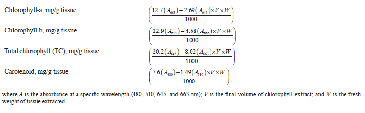
Estimation of chlorophylls and carotenoids using acetone. During this procedure, 1 g of finely cut fresh leaves was homogenized with 80% acetone. The mass was then centrifuged at 5000 rpm for 5 min. After the supernatant was transferred, the procedure was repeated until the residue contained no trace of green color. The final volume was brought up to 100 mL in the volumetric flask with 80% acetone. The optical density of the extracted solution was measured at 480, 510, 645, and 663 nm. From these readings, concentrations of chlorophylls and carotenoid pigment were determined by using the following formulas given in Table 1.
Gas chromatography–mass spectrometry (GC/MS). The aqueous extracts of C. sativum microgreens and mature leaves underwent a GC/MS analysis by using Agilent technologies 6890 N JEOL GC Mate II GC-MS model. The samples were injected into an HP-5 column (30 m×0.25 mm i.d with 0.25 μm film thickness). During the gas chromatography, helium served as the carrier gas, the flow rate was 1 mL/min, and the injector operated at 200°C. The column oven temperature was programmed as 50–250°C at a rate of 10°C/min injection mode. The list of mass spectrometry conditions included: ionization voltage – 70 eV; ion source temperature – 250°C; interface temperature – 250°C; mass range – 50–600 mass units. The results were compared using the spectrum of the known components stored in the National Institute Standard and Technology (NIST) library database [31].
![]()
The procedure made it possible to determine the sample concentration required to inhibit 50% of the DPPH free radical (IC50).
Ferric (Fe3+) reducing antioxidant power assay (FRAP). The reducing power of the extracts was determined by the Fe3+ reduction method with slight modification [33]. In brief, 1 mL of C. sativum extracts at different concentrations (50, 100, 150, 200, 250, and 300 μg/mL) were taken in 1 mL of phosphate buffer (0.2 M, pH 6.6) in a test tube. After that, 1 mL of potassium ferricyanide [K3Fe(CN)6] (1% w/v) was added. After 30 min of incubation at 50°C in a water bath, 1 mL of trichloroacetic acid (10 % w/v) was added to each mix. Then, 1 mL of fresh FeCl3(0.1% w/v) solution was poured in, and the absorbance was measured at 700 nm in a UV-Vis spectrophotometer. The experiment was replicated in three independent assays. Ascorbic acid was used as the standard reference. The reducing concentration (RC50) of sample required to reduce the free radicals (Fe3+) by 50 % was calculated to interpret the FRAP results.
The percentage of reduction was calculated as follows:
![]()
In vitroantidiabetic activity. α-amylase enzyme inhibition assay. The α-amylase enzyme inhibition assay relied on the starch-iodine test [34]. The coriander extracts at various concentrations (50, 100, 150, 200, 250, and 300 μg/mL) were added to α-amylase enzyme (10 μL). The α-amylase enzyme had been prepared in 0.02 M sodium phosphate buffer (pH 6.9 containing 6 mM sodium chloride). The procedure was followed by 10 min of incubation at 37°C. After pre-incubation, 500 μL of 1% soluble starch was added to each reaction and incubated at 37°C for 60 min.
To stop the enzymatic reaction, 1 N HCl (100 μL) was added and followed by 200 μL of iodine reagent (5 mM I2 and 5 mM KI). The color change was registered, and the optical density was tested at 595 nm. Acarbose was used as the standard reference. The control reaction representing 100% enzyme activity contained no plant extract.
The experiment was carried out in triplicate. A darkblue color indicated the presence of starch; a yellow color indicated the absence of starch; a brownish color indicated partially degraded starch in the reaction mix. In the presence of inhibitors, the starch added to the enzyme assay mix did not degrade and gave a darkblue color complex. No color complex developed in the absence of the inhibitor, indicating that starch was completely hydrolyzed by α-amylase. The IC50 value was calculated as follows:
![]()
Cytotoxicity assay on colon cell lines. The conventional MTT reduction assay was used to measure the cell viability [35]. HT 29 Colon cells were obtained from the National Centre for Cell Science (Pune). The culturing was performed on the medium developed by the Roswell Park Memorial Institute (RPMI). It included 10% fetal bovine serum (FBS), gentamycin (100 μg/mL), penicillin/streptomycin (250 U/mL), and amphotericin B (1 mg/mL). All cell cultures were maintained at 37°C in a humidified atmosphere of 5% CO2. Cells grew to confluence for 24 h before use.
As described in [36], we plated HT 29 cells (5×10³/ well) in 96-well plates for 24 h in 200 μL of the RPMI medium with 10% fetal bovine serum. After the culture supernatant was removed, the RPMI samples with various concentrations (0.001–100 μg/mL) of aqueous C. sativum extracts were added and incubated for 48 h. After the treatment, cells were incubated with MTT (10 μL, 5 mg/mL) at 37°C for 4 h and then with dimethyl sulfoxide at room temperature for 1 h. The plates were tested at 595 nm on a scanning multi-well spectrophotometer. All experiments were performed in duplicates [36].
The effect of the extracts on growth inhibition of HT-29 colon cancer cell line line, %, was calculated using the following formula:
![]()
From the above growth inhibition, (%) percentage of cell viability was derived using the following formula:
![]()
Statistical analysis. The phytochemical, antioxidant, and antidiabetic assays were carried out in triplicates, while the anticarcinogenic analysis was carried out in duplicates. The results obtained were expressed as mean ± SD. The statistical analysis was calculated by one-way ANOVA and Student’s t-test using Microsoft excel. All statistical significance was accepted at p < 0.05.
RESULTS AND DISCUSSION
Phytochemical analysis. Qualitative phytochemical analysis. The qualitative phytochemical analysis of the aqueous and ethanolic extracts of coriander microgreens and mature leaves revealed such phytochemicals as alkaloids, terpenoids, steroids, tannins, flavonoids, phenols, carbohydrates, and proteins. Saponins were absent in both aqueous and ethanol extracts of microgreens and mature leaves. However, glycosides were present in the aqueous extract of microgreens and mature leaves, as well as in the ethanol extract of mature leaves. However, they were absent in the ethanol extract of microgreens.
Quantitative phytochemical analysis. Tables 2 shows the quantitative phytochemical mean values of both aqueous and ethanol extracts of Coriandrum sativum microgreens and mature leaves.
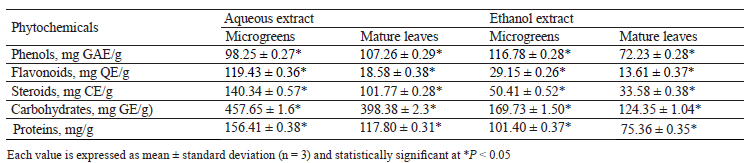
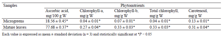
According to Table 2, the aqueous extract of microgreens showed a lower total phenol content (98.25 mg GAE/g) than that of mature leaves (107.26 mg GAE/g). However, the ethanol extract of microgreens had significantly (p < 0.05) higher total phenol (116.78 mg GAE/g) in comparison to that of mature leaves (72.23 mg GAE/g). In general, both extracts of microgreens had more total flavonoids, steroids, carbohydrates, and proteins than both extracts of mature leaves. Table 3 illustrates the contents of ascorbic acid, chlorophyll, and carotenoid.
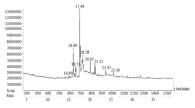
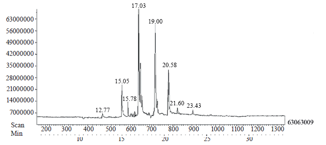
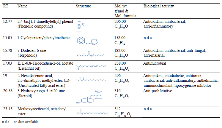
Gas chromatography–mass spectrometry (GC/MS). The GC/MS method revealed various bioactive constituents in the aqueous extracts of coriander microgreens and mature leaves. The analysis showed peaks at different locations on the chromatogram. In Figs. 1 and 2, the X-axis represents the retention time, while the Y-axis represents the relative abundance. The GC/MS analysis of a crude extract of microgreens showed nine major peaks. The crude extract of mature leaves eluted seven major peaks. Tables 4 and 5 illustrate a comparative analysis of the mass spectra of the constituents with the NIST library data.

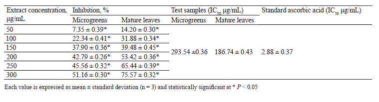
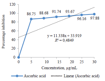
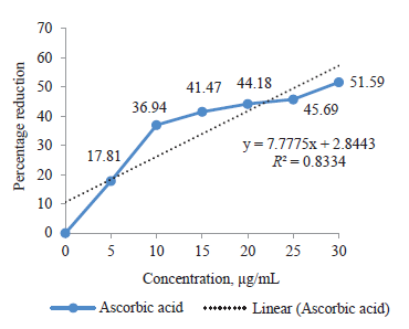
In vitro antioxidant assays. DPPH radical scavenging assay. The scavenging capacity of the aqueous and ethanol extracts of both coriander microgreens and mature leaves on DPPH free radicals was expressed as inhibition (%) (Tables 6 and 7). The IC50 was inhibition concentration at 50%: the lowest IC50 indicated the strongest ability of the extracts to act as DPPH radical scavengers. The aqueous and ethanol extracts of mature leaves showed the lowest IC50, which were 44.64 and 186.74 μg/mL, respectively. As for the aqueous and ethanol extracts of microgreens, they were 90.09 and 293.54 μg/mL, respectively. Compared to the reference standard ascorbic acid inhibition percentage (Fig. 3), the test samples required higher concentration to inhibit DPPH free radical. Thus, the test samples of microgreens and mature leaves showed dose-dependent scavenging activity.

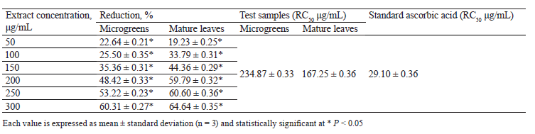
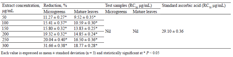
Ferric (Fe3+) reducing antioxidant power assay. For Fe3+ reducing activity, the ascorbic acid was used as standard. Figure 4 illustrates the standard curve; Tables 8 and 9 show the reducing power of test samples. The aqueous extract of mature leaves showed a slight increase in Fe3+ reduction compared to that of microgreens. The RC50 (50% reducing concentration) of microgreens and mature leaves in the aqueous extracts were 234.87 and 167.25 μg/mL, respectively. Interestingly, the ethanol extract of microgreens exhibited a greater ferric ion reducing power (31.66% at 300 μg/mL concentration) than that of mature leaves (18.77% at 300 μg/mL concentration). The ethanol extracts were unable to reduce the free radicals by RC50. The causes may be in some other chemical constituents that compete for reduction by Fe3+ and do not permit Fe3+ to donate an electron. The RC50 value for standard ascorbic acid was 29.1 μg/mL.
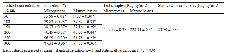
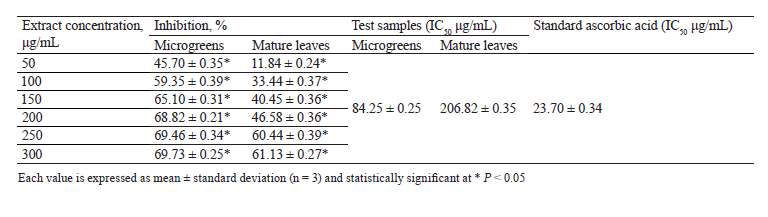
In vitro antidiabetic activity. α-amylase enzyme inhibition assay. Tables 10 and 11 show the inhibitory activity of test samples on the α-amylase enzyme. The aqueous and ethanol extracts of microgreens exhibited 50% of inhibition on α-amylase enzyme at 222.22 and 84.25 μg/mL. The results were lower than those of mature leaves IC50 values, which were 228.31 and 206.82 μg/mL, respectively. The standard reference drug acarbose (Fig. 5) showed α-amylase inhibitory activity with an IC50 valueof 23.71 μg/mL.

Cytotoxicity assay on colon cell lines. In the present study, the antioxidant activities of the aqueous extracts were compared to those of the ethanolic extracts. The aqueous extract of microgreens and mature leaves were examined for potential anticancer activity against the human colon HT-29 carcinoma cell line by using the MTT assay. The tests were performed in duplicate. The absorbance values were registered in the ELISA reader at 595 nm once purple color developed after 24 h of incubation. The mean was calculated for two trials (Table 12).
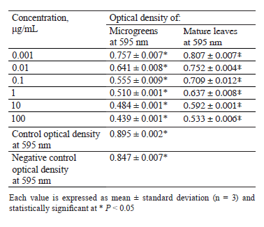
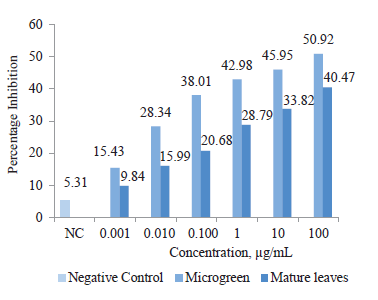
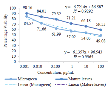
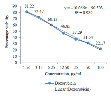
As the concentration of the test samples increased, the corresponding absorbance value decreased (p < 0.05). The MTT assay showed that the microgreen sample increased the percentage inhibition and consequently decreased the cell viability to 49.08% with the lowest IC50 value of 98.34 μg/mL. Mature leaves showed the least percentage inhibition and reduced viable cells to 59.53% with an IC50 value of 123.54 μg/mL (Figs. 6 and 7). Doxorubicin was used as the reference standard. Figure 8 demonstrates the standard curve of percent cell viability, which showed a cytotoxicity activity with an IC50 value of 11.75 μg/mL.
CONCLUSION
In the present research, aqueous and ethanol solvents of varying polarity were used to extract phytoconstituent compounds from Coriandrum sativum microgreens and mature leaves. The aqueous solvent had higher polarity for bio-extraction. According to the phytochemical analysis, C. sativum microgreens proved to be an abundant source of phenol, flavonoids, and steroids, compared to mature leaves. However, C. sativum mature leaves had more ascorbic acid, total chlorophylls, and carotenoids. The GC/MS test revealed various phytoconstituents with good therapeutic properties. The microgreens exhibited a much higher correlation with free radical reducing power than with the radical scavenging activity. The microgreens also had a higher α-amylase enzyme inhibitory property and a greater anticarcinogenic effecton colon cancer cell line. Therefore, C. sativum microgreens proved to be amore effective antioxidant, antidiabetic, and anticarcinogenic agent than mature leaves. Coriander microgreens can be as good as mature coriander leaves for the daily diet of a disease-free community.Contribution
The authors are equally involved in writing the manuscript and are equally responsible for plagiarism.CONFLICTS OF INTEREST
The authors have declared no conflict of interests regarding the publication of this manuscript.REFERENCES
- International Diabetes Federation [Internet]. [cited 2022 Jan 10]. Available from: https://www.idf.org/aboutdiabetes/what-is-diabetes/facts-figures.html
- González N, Prieto I, del Puerto-Nevado L, Portal-Nuñez, S, Ardura JA, Corton M, et al. 2017 update on the relationship between diabetes and colorectal cancer: Epidemiology, potential molecular mechanisms and therapeutic implications. Oncotarget. 2017;8(11):18456–18485. https://doi.org/10.18632/oncotarget.14472
- Suh S, Kim K-W. Diabetes and cancer: is diabetes causally related to cancer? Diabetes and Metabolism Journal. 2011;35(3)193–198. https://doi.org/10.4093/dmj.2011.35.3.193
- Meyer B, Are C. Current status and future directions in colorectal cancer. Indian Journal of Surgical Oncology. 2018;9(4):440–441. https://doi.org/10.1007/s13193-017-0711-9
- Nishteswar K. Ayurvedic concept of food and nutrition. Journal of Nutrition & Food Sciences. 2017;6(4). https://doi.org/10.4172/2155-9600.1000530
- De Souza EL, Stamford TLM, De Oliveira Lima E, Tarajano VN, Barbosa Filho JM. Antimicrobial effectiveness of spices: An approach for use in food conservation system. Brazilian Archives Biology Technology. 2005;48(4):549–558. https://doi.org/10.1590/s1516-89132005000500007
- Sachan AKR, Kumar S, Kumari K, Singh D. Medicinal uses of spices used in our traditional culture: World wide. Journal of Medicinal Plants Studies. 2018;6(3):116–122.
- Prachayasittikul V, Prachayasittikul S, Ruchirawat S, Prachayasittikul V. Coriander (Coriandrum sativum): A promising functional food toward the well-being. Food Research International. 2018;105:305–323. https://doi.org/10.1016/j.foodres.2017.11.019
- Pinto E, Almeida AA, Aguiar AA, Ferreira IMPLVO. Comparison between the mineral profile and nitrate content of microgreens and mature lettuces. Journal of Food Composition and Analysis. 2015;37:38–43. https://doi.org/10.1016/j.jfca.2014.06.018
- Xiao Z, Lester GE, Luo Y, Wang Q. Assessment of vitamin and carotenoid concentrations of emerging food products: Edible microgreens. Journal of Agricultural and Food Chemistry. 2012;60(31):7644–7651. https://doi.org/10.1021/jf300459b
- Ebert AW, Wu TH, Yang RY. Amaranth sprouts and microgreens-A homestead vegetable production option to enhance food and nutrition security in the rural-urban continuum. SEAVEG2014: families, farms, food: Proceedings of the regional symposium on sustaining small-scale vegetable production and marketing systems for food and nutrition security; 2014; Bangkok. Taiwan: AVRDC; 2015. p. 233–244.
- Kou L, Yang T, Luo Y, Liu X, Huang L, Codling E. Pre-harvest calcium application increases biomass and delays senescence of broccoli microgreens. Postharvest Biology and Technology. 2014;87:70–78. https://doi.org/10.1016/j.postharvbio.2013.08.004
- Mir SA, Shah MA, Mir MM. Microgreens: Production, shelf life and bioactive components. Critical Reviews in Food Science and Nutrition. 2017;57(12):2730–2736. https://doi.org/10.1080/10408398.2016.1144557
- Chandra D, Kim JG, Kim YP. Changes in microbial population and quality of microgreens treated with different sanitizers and packaging films. Horticulture Environment and Biotechnology. 2012;53(1):32–40. https://doi.org/10.1007/s13580-012-0075-6
- Banu SK, Cathrine L. General techniques involved in phytochemical analysis. International Journal of Advanced Research in Chemical Science. 2015;2(4):25–32.
- Finar IL. Stereo chemistry and the chemistry of natural products. Vol. 2. Longman; 1986. 518 p.
- Hindi NKK, Al-Charrakh AH, Naher HS, Abbas AS. Study of chemical analysis of Iraqi propolis and active component of proplis, Iraq. Journal of Science. 2015;15(11):1095–1103.
- Kokate CK. Practical pharmacognosy. 4th edition. New Delhi: Vallabh Prakashan; 1994. 186 p.
- Shrivastava DK. Phytochemical analysis of a miracle herb Coriander sativum. Indian Journal of Scientific Research. 2017;13(2):09–14.
- Singh V, Kumar R. Study of phytochemical analysis and antioxidant activity of Allium sativum of Bundelkhand region. International Journal of Life Sciences Scientific Research. 2017;3(6):1451–1458.
- Wagner H, Bladt S. Plant drug analysis. A thin layer chromatography atlas. Heidelberg: Springer Berlin; 1996. 384 p. https://doi.org/10.1007/978-3-642-00574-9
- Singleton VL, Orthofer R, Lamuela-Raventós RM. Analysis of total phenols and other oxidation substrates and antioxidants by means of folin-ciocalteu reagent. Methods in Enzymology. 1999;299:152–178. https://doi.org/10.1016/S0076-6879(99)99017-1
- Ahmed D, Fatima K, Saeed R. Analysis of phenolic and flavonoid contents, and the anti-oxidative potential and lipid peroxidation inhibitory activity of methanolic extract of Carissa opaca roots and its fractions in different solvents. Antioxidants. 2014;3(4):671–683. https://doi.org/10.3390/antiox3040671
- Kharate MS, Pandhure NB. Quantitative estimation of phytochemical and antimicrobial properties of crude leaf extract, Mimusops elengil L. International journal for research in applied science & engineering technology. 2017;5(12):2699–2707.
- Hedge JE, Hofreiter BT. Carbohydrate chemistry. 17th edition. New York: Academic Press; 1962.
- Mahavi MO, Nilesh PS. Quantitative estimation of amino acids and carbohydrates in the root exudates of Salvadora persica. International Journal of Biology Research. 2016;1(3):4–10.
- Rao BS, Deshpande V. Experimental biochemistry: A student companion. Royal Tunbridge Wells: Anshan; 2006. 302 p.
- Aron DI. Copper enzymes in isolated chloroplasts phenoloxidase in Beta vulgaris. Plant physiology. 1949;24(1):1–15. https://doi.org/10.1104/pp.24.1.1
- Duxbury AC, Yentach CS. Plankton pigment monograph. Journal of Marine Research. 1956;15:190–191.
- Maclachalam S, Zalik S. Plastid structure, chlorophyll concentration and free amino acid composition of a chlorophyll mutant of barely. Canadian Science Publishing. 1963;4:1053–1062.
- Ravisankar N, Sivaraj C, Seeni S, Joseph J, Raaman N. GC-MS Analysis and anticancer activity of methanol extract of leaves of Hypericum hookerianum Wight & Arn. International Journal of Pharmacy and Pharmaceutical Sciences. 2014;6(5):515–519.
- Khalaf NA, Shakya AK, Al-othman A, El-agbar Z, Farah H. Antioxidant activity of some common plant. Turkish Journal of Biology. 2008;32(1):51–55.
- Oyaizu M. Studies on products of browning reaction: antioxidative activities of products of browning reaction prepared from glucosamine. Japanese Journal of Nutrition. 1986;44(4):307–315. https://doi.org/10.5264/eiyogakuzashi.44.307
- Gayathri S, Sivaraj C, Sangeetha ST, Arumugam P, Manimaran A. Evaluation of antioxidant, antibacterial, alpha amylase enzyme inhibition activities and GC-MS analysis of leaves extract of Gymnema sylvestre L. Journal of Pharmacognosy and Phytochemistry. 2018;7(6):2326–2333.
- Mosmann T. Rapid colorimetric assay for cellular growth and survival: Application to proliferation and cytotoxicity assays. Journal of Immunological Methods. 1983;65(1–2):55–63. https://doi.org/10.1016/0022-1759(83)90303-4
- Vardhini SP, Sivaraj C, Arumugam P, Himanshu R, Kumaran T, Baskar M. Antioxidant, anticancer, antibacterial activities and GCMS analysis of aqueous extract of pulps of Aegle marmelos (L.) Correa. The Journal of Phytopharmacology. 2018;7(1):72–78. https://doi.org/10.31254/phyto.2018.7115
