

Abstract
The awareness of some harmful side effects of the chemicals contained in synthetic cosmetics has increased the demand for herbal-based cosmetic products today.White pitahaya fruit and peel methanol extracts were prepared to determine their usage potential in the cosmetic industry. Firstly, we investigated their antimicrobial activity against some test microorganisms using the disc diffusion assay. We also determined their minimal inhibition and minimal bactericidal or fungicidal concentrations. Then, we assayed the antimicrobial activity of a commercial cream containing white pitahaya extracts and the probiotic Lactobacillus fermentum MA-7 strain against the test microorganisms. Finally, we measured the sun protection factors of the white pitahaya fruit and peel extracts and the cream with the extracts.
The white pitahaya fruit and peel extracts exhibited antimicrobial activity against the test microorganisms. The cream formulation containing a pitahaya fruit extract had the highest inhibition zone diameter of 11.25 mm against Escherichia coli O157:H7. The highest sun protection value among the extracts and cream with extracts was determined for peel extract as 6.66 and 23.34, respectively.
The results indicate that pitahaya fruit and peel extracts have effective antibacterial and antifungal properties, as well as high sun protection factors, and therefore they could be used as natural preservatives in the cosmetic industry.
Keywords
Extract, antimicrobials, probiotics, cream formulation, solar protectionINTRODUCTION
Pitahaya is a tropical fruit belonging to the Cactaceae family which attracts the attention of researchers and producers due to its appearance and color [1]. It was first used as an ornamental plant, and then its fresh fruit began to be consumed [2]. Pitahaya fruit is rich in vitamins and fiber, which helps the digestive system, blocks diabetes, removes toxic substances such as heavy metals, as well as controls cholesterol and blood pressure levels [2]. Pitahaya is known to have a high content of antioxidants which help fight many diseases such as arthritis, atherosclerosis, cancer, heart disease, inflammation, and brain dysfunction [3].
Food-borne and clinical pathogens pose a significant threat to human health. In developed countries, these pathogens cause millions of infectious gastrointestinal diseases each year [4]. The influence of fungal microorganisms on human health has been increasing rapidly in recent years, with the global mortality rate from fungal diseases exceeding that from malaria or breast cancer [5]. Chemical-containing substances can be used against these pathogens, but they have harmful effects on human health, such as resistance to antimicrobials or toxicity.
Therefore, there is a growing interest in finding safer and more effective natural preservatives. Plants are natural sources of antimicrobials that can be used against various infectious pathogens, such as bacteria and fungi. More than 1340 plants with antimicrobial activity have been identified and more than 30 000 natural antimicrobial compounds have been isolated [6].
Formulations containing probiotics are increasingly in demand for skin care, prevention and treatment of skin aging and skin diseases. Thus, the use of probiotics in the cosmetic industry represents an emerging area for skin health [7]. Roudsari et al. reported that probiotics in cosmetic formulations have protective effects against atopic dermatitis, acne, eczema, allergic inflammation, or hypersensitivity of the skin to ultraviolet-induced damage [8]. The skin, one of the important organs of the human body, acts as a protective wall that prevents disease-causing pathogenic microorganisms from entering the internal system of the body.
It has been reported in many studies, e.g. by Nichols and Katiyar, that prolonged exposure to sunlight causes various skin conditions [9]. Solar UV rays are divided into three regions: UV-A (320–400 nm), UV-B (290– 320 nm), and UV-C (200–290 nm). UV-A rays cause serious problems such as suppression of the immune system, premature skin aging, and endothelial cell necrosis that damages the dermal blood vessels [10]. UV-B rays cause sunburn, suppression of the immune system, photo carcinogenesis, and pigmentation [11]. UV-C rays react with oxygen atoms in the atmosphere and convert oxygen into ozone, which is absorbed by the atmosphere before reaching the Earth [12]. Sunscreen creams are used to protect the skin against the damage of these rays. However, chemical sunscreen creams can have some harmful effects on people with sensitive skin. Recent years have seen a trend towards using natural products to reduce or eliminate these harmful effects. In particular, researchers have focused on the development of herbal sunscreens that have no side effects by including naturally produced bioactive substances into sunscreens [10].
Fruit extracts are known to have ultraviolet (UV) protection, as well as antimicrobial and antioxidant activities due to the compounds they contain [13, 14]. Therefore, plant extracts have the potential to be used as bioactive natural products in food preservation, pharmaceutical industry, healthcare, and cosmetics [15].
We aimed to 1) evaluate the antimicrobial activity of pitahaya extracts; 2) develop a cream formulation containing pitahaya as a natural ingredient; 3) measure the sun protection factors of the pitahaya extracts to determine their potential use in the cosmetic industry as an additive in natural sunscreens; and 4) investigate the solar protection activity of the commercial cream with pitahaya extracts.
STUDY OBJECTS AND METHODS
Plant material. The white pitahaya fruit was purchased in Antalya (Turkey) in September 2021.
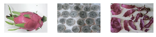
Preparation of extracts. The pitahaya fruit samples were washed with distilled water. Then, the fruits and peels were air-dried separately at room temperature (Fig. 1). The dried fruit samples were ground with a Waring blender. For extraction, 10 g of the plant material was mixed with 30 mL of methanol (96%). The fruit samples were extracted every day for 6 h (3 days) in a hot water bath. The extracts were concentrated by using a rotary evaporator. The white pitahaya fruit and peel extracts were dissolved in dimethyl sulfoxide (DMSO) and then sterilized with a 0.45-μm filter. The extracts were stored at 4°C until used.
Determination of antimicrobial activity. Disc diffusion assay. The disc diffusion method was used to determine the antimicrobial activity of the methanol extracts from white pitahaya fruit and peel against test microorganisms, including food-borne and clinical bacteria (Salmonella enteritidis RSKK 171, Pseudomonas aeruginosa ATCC 27853, Listeria monocytogenes ATCC 7644, Enterococcus faecalis ATCC 29212, and Escherichia coli O157:H7) and two yeasts (Candida albicans ATCC 10231 and Candida glabrata RSKK 04019).
The food-borne and clinical pathogens were cultured at 37°C in Tryptic Soy Broth and Nutrient Broth media, while the yeasts were cultured at 30°C in Yeast Peptone Dextrose media for 24 h. The test microorganisms were washed twice with a saline solution and their concentration was adjusted to 0.5 McFarland. 100 μL of the prepared microbial suspension (0.5 McFarland) was spread onto agar media. Sterile discs (6 mm in diameter) were placed on the solid agar and 20 μL (4000 μg/disc) of the white pitahaya fruit and peel extracts were then dropped onto the discs in triplicates. The petri dishes were incubated at appropriate temperatures for 24 h. After incubation, the inhibition zones around the discs were measured with a caliper and recorded.
Micro-dilution assay. The minimum inhibition concentration and minimum bactericidal concentration or minimum fungicidal concentration of the extracts were determined using the micro-dilution assay. Test microorganisms adjusted to 0.5 McFarland were added to each tube containing the extract and the media. The mixtures in the tubes were incubated under the same conditions as in the disc diffusion assay for each microorganism. After incubation, the concentration of the extract in the non-growth tube was recorded as minimum inhibition concentration values. Then, the samples from the tubes were inoculated onto solid media using the spot-dropping method. The petri dishes were incubated for 24 h at the same temperature for each microorganism which is indicated in the minimum inhibition concentration determination assay. At the end of the incubation, the extract concentrations that prevented bacterial growth on the solid media were evaluated as minimum bactericidal concentration or minimum fungicidal concentration values.
Antimicrobial activity of the cream with pitahaya extract and probiotic. A new method was designed by modifying the protocols of Handali et al. and Chen et al. [16, 17]. To formulate an antimicrobial cream, we used a commercial cream, white pitahaya fruit extracts, and the Lactobacillus fermentum MA-7 probiotic strain isolated from human milk [18].
In this study, three different experimental samples were assayed. The control was the cream (10% w/v) adjusted with distilled water to the final volume. The experimental samples contained: a) the cream (10% w/v) with the L. fermentum MA-7 probiotic strain; b) the cream (10% w/v) with the white pitahaya extract (20% w/v); and c) the cream (10% w/v) with the white pitahaya extract (20% w/v) and the L. fermentum MA-7 strain.
After vortexing, all the groups were sonicated for 15 min and then sterilized with a 0.45-μm filter. The antimicrobial activity of the prepared mixtures was determined by the well diffusion method. The test microorganisms used in this study included S. enteritidis RSKK 171, E. faecalis ATCC 29212, P. aeruginosa ATCC 27853, E. coli O157:H7, L. monocytogenes ATCC 7644, C. albicans ATCC 10231, and C. glabrata RSKK 04019. They were washed twice with a saline solution and their concentrations were adjusted to 0.5 McFarland. The microorganism suspensions (100 μL) were spread onto solid media. 100 μL of the mixture was added to each well (6 mm in diameter). The volume of the wells corresponded to 0.1. The experiment was carried out in three repetitions. The petri dishes were incubated for 24 h at 37°C for the food-borne and clinical pathogens and at 30°C for the yeasts. The inhibition zones around the wells were then measured with a caliper and recorded.
Sun protection factor of pitahaya extract. The sun protection factor (SPF) of the white pitahaya fruit and peel extracts was determined spectrophotometrically. The extract (0.006 g) was mixed with 3 mL of ethanol (96%) and homogenized. The homogeneous mixture was measured using a spectrophotometer (Beckman Coulter, USA) at 5-nm intervals in the wavelength range of 290– 320 nm. The recorded values were calculated using the Mansur equation [19]:
![]()
where CF is the correction factor (= 10); EE(λ) is the erythemetogenic effect radiation wavelength (λ); I(λ) is the intensity of sunlight at wavelength (λ); Abs(λ) is the absorbance of extracts at wavelength (λ).
Sun protection factor of the pitahaya extract and cream samples. The sun protection factor of the pitahaya extract and commercial cream samples was determined using a modified method of Bambal et al. and Imam et al. [20, 21]. For this, 1 g of cream and 0.5 g of white pitahaya (fruit and peel) extracts were mixed, and 8.5 g of distilled water was added to the mixture. The mixture was stirred until it became homogeneous. 0.1 g of the homogeneous mixture was taken into another tube and completed to 10 mL volume with ethanol (40%). After sonication (30 kHz, 100% amplitude) for 5 min, the mixture was filtered through Whatman No. 1 filter paper. The mixture (0.5 mL) was adjusted to 5.0 mL with ethanol (40%) in another tube. Then, 0.5 mL of this mixture was taken, and the volume was completed with ethanol (40%) to 2.5 mL. The mixtures adjusted to 2.5, 5.0, and 10.0 mL were measured in three repetitions using the spectrophotometer (Beckman Coulter, USA) at 5-nm intervals in the wavelength range of 290–320 nm. The sun protection factor values of the control and experimental samples were calculated using the Mansur equation presented above [19].
RESULTS AND DISCUSSION
We investigated the antimicrobial and antifungal activities of the white pitahaya fruit and peel extracts on food-borne and clinical test microorganisms. Disc diffusion assay results are given in Table 1. As we can see, the highest inhibition zone diameter (11.57 mm) was found against Pseudomonas aeruginosa ATCC 2785 in the peel extract, while the lowest inhibition zone diameter (6.17 mm) was found against Escherichia coli O157:H7 in the fruit extract. As for the antifungal activity, the fruit and peel extracts had inhibition zone diameters of 11.66 and 13.15 mm, respectively, against Candida albicans ATCC 10231 and 12.21 and 12.93 mm, respectively, against Candida glabrata RSKK 04019.
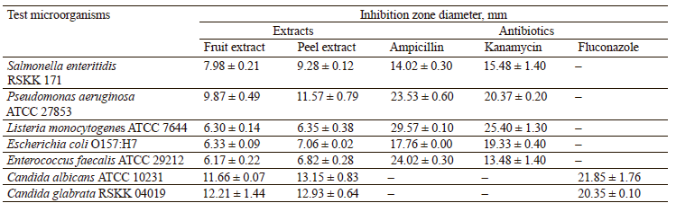
Nurmahani et al. stated that ethanol, hexane, and chloroform extracts of white pitahaya peel showed an inhibition zone diameter of 7–9 mm against Bacillus cereus ATCC 14579, Staphylococcus aureus ATCC 25923, Listeria monocytogenes ATCC 19115, Enterococcus faecalis ATCC 14506, Salmonella typhimurium ATCC 13311, E. coli ATCC 25922, Klebsiella pneumoniae ATCC 13883, Yersinia enterocolitica ATCC 23715, and Campylobacter jejuni ATCC 29428 [22]. In our study, the inhibition zone diameters were larger compared to their results. In another study by Mahdi et al., who used the well diffusion method, the inhibition zone diameters of 7–11 mm were obtained for white pitahaya aqueous extracts against E. coli, Klebsiella spp., Staphylococcus epidermidis, S. aureus, and C. albicans [23]. The differences in the antimicrobial activity assay results may be due to the differences in the origin of the pitahaya fruit samples, the solvents, as well as extraction and test methods used in the studies.
The disc diffusion assay results indicated the white pitahaya extracts’ antimicrobial or antifungal activity against all the tested microorganisms. The minimum inhibition concentration, minimum bactericidal concentration or minimum fungicidal concentration values are shown in Table 2.
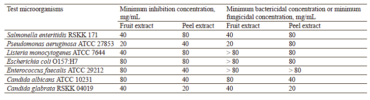
Among the tested white pitahaya extracts, the lowest minimum bactericidal concentration value was found to be 20 mg/mL for the fruit extract against P. aeruginosa ATCC 27853. The minimum bactericidal concentration values of the fruit extracts varied from 20 to ≥ 80 mg/mL. The minimum bactericidal concentration values for the peel extracts were found to be 80 or ≥ 80 mg/mL against the tested bacterial strains. The lowest minimum fungicidal concentration value was 20 mg/mL for the peel extract against C. glabrata RSKK 04019.
The tested extract was considered bactericidal if the minimum bactericidal/inhibition concentration ratio was ≤ 4 and bacteriostatic if the minimum bactericidal/ inhibition concentration ratio was > 4 [24, 25]. The minimum bactericidal/inhibition concentration or minimum fungicidal/inhibition concentration ratios of some white pitahaya fruit and peel extracts were found to be 1 and 2. We established that those extracts had antibacterial and antifungal activities against the tested microorganisms, except for the not determined group (Table 3).

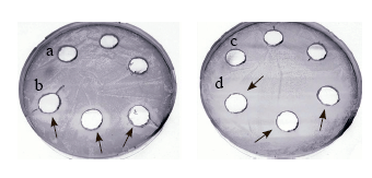
The antimicrobial activity assay results of the cream samples are given in Table 4. As we can see, the cream with the extract and Lactobacillus fermentum formed an inhibition zone against all the tested microorganisms. The highest inhibition zone diameter (11.25 mm) among the tested microorganisms was obtained against E. coli O157:H7 in the cream with the extract and L. fermentum. The control cream and the cream with the extract did not show an inhibition zone against E. coli O157:H7, while the cream with L. fermentum mixture had a diameter of 6.4 mm.
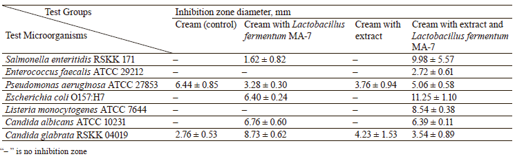
According to Fig. 2, the antimicrobial activity or increased inhibition zone diameters were observed in the cream with the extract and L. fermentum, compared to the control sample. The results for the control and all the test groups showed that the pitahaya extract and the probiotic candidate strain increased the antimicrobial activity of the commercial cream by creating a synergetic effect.
A recent study by Moysidis et al. indicated that the cream sample with L. fermentum MA-7 increased the rate of healing the wound [26]. Another recent study showed inhibition zone diameters of 2–10 mm in cream with Vernonia ambigua leave extracts against C. albicans, S. aureus, E. coli, and P. aeruginosa [27].
Sunscreens, which are widely used today to protect the skin from sun rays, constitute a large part of the cosmetic industry. In our study, the white pitahaya fruit and peel extracts had a sun protection factor of 3.95 and 6.66, respectively (Fig. 3). According to the UV blocking rates reported by Imam et al., the white pitahaya fruit and peel extracts had a UV blocking capacity of 75 and 80%, respectively [21].

The sun protection factor values of the cream samples containing white pitahaya fruit and peel extracts are given in Fig. 4. The highest sun protection factor values of 9.26 and 23.34 were observed in the fruit and peel extracts at 10-mL concentrations, respectively. The highest sun protection factor value was observed at the 10-mL concentration for the samples with the extract as 9.26 and 23.34, while that for the commercial cream (control) was found to be 4.94 (Fig. 4). According to Imam et al., the cream samples with pitahaya fruit and peel extracts had UV blocking rates of 90 and 95%, respectively [21]. Compared to the control, the fruit and peel extracts increased the sun protection factor values and the UV blocking rate of the cream.
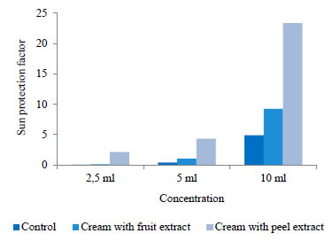
Higher concentrations of the extract added to the commercial cream increased the sun protection factor value of the cream. The sun protective properties of plant extracts are due to the phenolic compounds and flavonoid substances they contain. Phenolic and flavonoid compounds found in pitahaya have been reported to contribute to a high sun protection factor value and broad-spectrum UV-A and UV-B light protection [28]. The use of natural additives obtained from plants in various industries enables low-cost production in high quantities [29, 30].
CONCLUSION
We studied the biological activities of pitahaya fruit and peel extracts to determine their usage potential in the cosmetic industry. The extracts exhibited good antibacterial and antifungal activities against the tested microorganisms. We also tested the cream samples containing the pitahaya extracts to determine their antimicrobial activity. The cream with the pitahaya fruit extract and Lactobacillus fermentum MA-7 with high antimicrobial activity could become an alternative to chemical preservatives. Additionally, the pitahaya fruit extracts and the cream with the pitahaya extracts exhibited high sun protection values. The results showed that pitahaya fruit and peel extracts could be suitable as natural preservatives for the cosmetic industry.Contribution
The authors were equally involved in writing the manuscript and are equally responsible for plagiarism.CONFLICTS OF INTEREST
The authors declare that they have no conflict of interest.FUNDING
This research received no specific grant from any funding agency in the public, commercial, or not-for-profit sectors.REFERENCES
- Panisson D, Marques NK, de Souza FBM, Neto JCM, Freire AI, de Araújo NO, et al. Growth and initial development of pitaya white (Hylocereus undatus) and red (Hylocereus monacanthus) in the city of Araguaína-TO. Research, Society and Development. 2021;10(14). https://doi.org/10.33448/rsd-v10i14.21921
- Gunasena HPM, Pushpakumara DKNG, Kariyawasam M. Dragon fruit: Hylocereus undatus (Haw.) Britton and Rose. In: Pushpakumara DKNG, Gunasena HPM, Singh VP, editors. Underutilized fruit trees in Sri Lanka., New Delhi: World Agroforestry Centre, South Asia Office; 2006. pp. 110–141.
- Susanti EVH, Budi Utomo S, Syukri Y, Redjeki T. Phytochemical screening and analysis polyphenolic antioxidant of methanolic extract of white dragon fruit (Hylocereus undatus). Indonesian Journal of Pharmacy. 2012;23(1):60–64.
- Jin S-Q, Yin B-C, Ye B-C. Multiplexed bead-based mesofluidic system for detection of food-borne pathogenic bacteria. Applied and Environmental Microbiology. 2009;75(21):6647 –6654. https://doi.org/10.1128/AEM.00854-09
- Brown GD, Denning DW, Gow NAR, Levitz SM, Netea MG, White TC. Hidden killers: Human fungal infections Science Translational Medicine. 2012;4(165). https://doi.org/10.1126/scitranslmed.3004404
- Tajkarimi MM, Ibrahim SA, Cliver DO. Antimicrobial herb and spice compounds in food. Food Control. 2010;21(9):1199–1218. https://doi.org/10.1016/j.foodcont.2010.02.003
- Cinque B, Torre CL, Melchiorre E, Marchesani G, Zoccali G, Palumbo P, et al. Use of probiotics for dermal applications. In: Liong M-T, editor. Probiotics. Biology, genetics and health aspects. Heidelberg: Springer Berlin; 2011. pp. 221–241. https://doi.org/10.1007/978-3-642-20838-6_9
- Roudsari MR, Karimi R, Sohrabvandi S, Mortazavian AM. Health effects of probiotics on the skin. Critical Reviews in Food Science and Nutrition. 2015;55(9):1219–1240. https://doi.org/10.1080/10408398.2012.680078
- Nichols JA, Katiyar SK. Skin photoprotection by natural polyphenols: Anti-inflammatory, antioxidant, and DNA repair mechanisms. Archives of Dermatological Research. 2010;302(2):71–83. https://doi.org/10.1007/s00403-009-1001-3
- Mishra AK, Mishra A, Chattopadhyay P. Herbal cosmeceuticals for photoprotection from ultraviolet B radiation: A review. Tropical Journal of Pharmaceutical Research. 2011;10(3):351–360. https://doi.org/10.4314/tjpr.v10i3.7
- DeBuys HV, Levy SB, Murray JC, Madey DL, Pinnell SR. Modern approaches to photoprotection. Dermatological Clinics. 2000;18(4):577–590. https://doi.org/10.1016/S0733-8635(05)70208-4
- Dutra EA, Da Costa E Oliveira, DAG, Kedor-Hackmann ERM, Miritello Santoro MIR. Determination of sun protection factor (SPF) of sunscreens by ultraviolet spectrophotometry. Brazilian Journal of Pharmaceutical Sciences. 2004;40(3):381–385. https://doi.org/10.1590/S1516-93322004000300014
- Ahmad I, Beg AZ. Antimicrobial and phytochemical studies on 45 Indian medicinal plants against multi-drug resistant human pathogens. Journal of Ethnopharmacology. 2001;74(2):113–123. https://doi.org/10.1016/S0378-8741(00)00335-4
- Cushnie TPT, Lamb AJ. Antimicrobial activity of flavonoids. International Journal of Antimicrobial Agents. 2005;26(5):343–356. https://doi.org/10.1016/j.ijantimicag.2005.09.002
- Sakhr K, El Khatib S. Physiochemical properties, and medicinal, nutritional, and industrial applications of Lebanese Sumac (Syrian Sumac – Rhus coriaria): A review. Heliyon. 2020;6(1). https://doi.org/10.1016/j.heliyon.2020.e03207
- Handali S, Hosseini H, Ameri A, Moghimipour E. Formulation and evaluation of an antibacterial cream from Oxalis corniculata aqueous extract. Jundishapur Journal of Microbiology. 2011;4(4):255–260.
- Chen MX, Alexander KS, Baki G. Formulation and evaluation of antibacterial creams and gels containing metal ions for topical application. Journal of Pharmaceutics. 2016;2016. https://doi.org/10.1155/2016/5754349
- Asan-Ozusaglam M, Gunyakti A. Lactobacillus fermentum strains from human breast milk with probiotic properties and cholesterol-lowering effects. Food Science and Biotechnology. 2019;28(2):501–509. https://doi.org/10.1007/s10068-018-0494-y
- Mansur JDS, Breder MNR, Mansur MCA, Azulay RD. Correlation between the determination of sun protecting of factor in human beings and by spectrophotometry. Anais Brasileiros de Dermatologia. 1986;61(4):167–172.
- Bambal V, Wyawahare N, Turaskar A, Mishra M. Study of sunscreen activity of herbal cream containing flower extract of Nyctanthes arbortristis L. and Tagetes erecta L. International Journal of Pharmaceutical Sciences Review and Research. 2011;11(1):142–146.
- Imam S, Azhar IN, Mahmood ZA. In vitro evaluation of sun protection factor of a cream formulation prepared from extracts of Musa accuminata (L.), Psidium gujava (L.) and Pyrus communis (L.). Asian Journal of Pharmaceutical and Clinical Research. 2015;8(3):234–237.
- Nurmahani MM, Osman A, Abdul Hamid A, Mohamad Ghazali F, Pak Dek MS. Antibacterial property of peel extracts of Hylocereus polyrhizus and Hylocereus undatus. International Food Research Journal. 2012;19(1):77–84.
- Mahdi MA, Mohammed MT, Jassim AMN, Mohammed AI. Phytochemical content and anti-oxidant activity of Hylocereus undatus and study of toxicity and the ability of wound treatment. Plant Archives. 2018;18(2):2672–2680.
- Gatsing D, Tchakoute V, Ngamga D, Kuiate J-R, Tamokou JDD, Nji-Nkah BF, et al. In vitro antibacterial activity of Crinum purpurascens Herb. leaf extract against the Salmonella species causing typhoid fever and its toxicological evaluation. Iranian Journal of Medical Sciences. 2009;34(2):126–136.
- Hazen KC. Fungicidal versus fungistatic activity of terbinafine and itraconazole: An in vitro comparison. Journal of the American Academy of Dermatology. 1998;38(5):S37–S41. https://doi.org/10.1016/S0190-9622(98)70482-7
- Moysidis M, Stayrou G, Cheva A, Abba Deka A, Tsetis JK, Birba V, et al. The 3-D configuration of excisional skin wound healing after topical probiotic application. Injury. 2022;53(4):1385–1393. https://doi.org/10.1016/j.injury.2022.02.006
- Okafo SE, Anie CO, Nwanua MC. Formulation and evaluation of antimicrobial topical creams from ethanol extract of Vernonia ambigua leaves. Nigerian Journal of Pharmaceutical Research. 2019;15(2):249–255. https://doi.org/10.4314/njpr.v15i2.12
- Vijayakumar R, Abd Gani SS, Zaidan UH, Halmi MIE, Karunakaran T, Hamdan MR. Exploring the potential use of Hylocereus polyrhizus peels as a source of cosmeceutical sunscreen agent for its antioxidant and photoprotective properties. Evidence-Based Complementary and Alternative Medicine. 2020;2020. https://doi.org/10.1155/2020/7520736
- Farasat M, Khavari-Nejad R-A, Nabavi SMB, Namjooyan F. Antioxidant activity, total phenolics and flavonoid contents of some edible green seaweeds from northern coasts of the Persian gulf. Iranian Journal of Pharmaceutical Research. 2014;13(1):163–170.
- Mirshafa S-A, Azadbakht M, Ahangar N. Study of antidepressant and sedative-hypnotic activity of hydroalcoholic extract of Asperugo procumbens L. aerial parts in mice. Iranian Journal of Pharmaceutical Research. 2013;12(3):529–535.
