

Аннотация
Introduction. Osteoporosis is one of the most common diseases of the musculoskeletal system in modern clinical practice. Its prevention and treatment requires a diet with a sufficient intake of calcium, vitamins, and connective tissue proteins that regenerate cartilage and bone tissue. We aimed to formulate a functional product based on collagen fermentolysate to prevent osteoporosis and prove its effects in experiments on laboratory rats.Study objects and methods. Our study objects were collagen fermentolysate obtained from pork ears and legs (1:1) and the functional product based on it. The biological experiment was carried out on Wistar female rats exposed to osteoporosis through complete ovariectomy. Their femurs were analyzed for the contents of phosphorus, magnesium, and calcium, as well as cytometric and biochemical blood parameters.
Results and discussion. The formulated functional product based on collagen fermentolysate contained 41% of the most easily assimilable peptide fractions with a low molecular weight of 10 to 20 kDa. Other components included pumpkin powder, dietary fiber, calcium, chondroprotectors, and vitamins E, C, and D3. Compared to the control, the experimental rats that received the functional product had increased contents of calcium and magnesium in the bone tissue (by 25.0 and 3.0%, respectively), a decreased content of phosphorus (by 7.0%), a calcium-to-phosphorus ratio restored to 2.4:1.0, and a higher concentration of osteocalcin in the blood serum (by 15%).
Conclusion. The developed functional product based on collagen fermentolysate can be used as an additional source of connective tissue protein, calcium, vitamins C, E, and D3, dietary fiber, and chondroprotectors to prevent osteoporosis.
Ключевые слова
Collagen, fermentolysate, osteoporosis, functional foods, raw meat, calcium, oophorectomyВВЕДЕНИЕ
Current global trends in food production are aimed at designing healthy foods to improve public health and prevent diseases caused by unbalanced nutrition. In the recent years, the quality and safety of food products has been a strategic priority in Russia. New laws have been passed to regulate and encourage the development and production of a wide range of healthy foods, including functional products.
Diseases of the musculoskeletal system are among the most common in modern clinical practice, especially osteoporosis. According to the World Health Organization, almost 200 million people suffer from osteoporosis worldwide, with over 9 million fractures occurring every year. Women aged 55+ are especially vulnerable to this pathology, which is presumably associated with estrogen deficiency in the postmenopausal period.
Postmenopausal osteoporosis is caused by accelerated bone resorption and systemic calcium imbalance. Osteoporosis caused by hypoestrogenism is commonly treated with drugs that prevent bone resorption or stimulate the formation of bone tissue. These drugs are mainly based on female sex hormones or selective estrogen receptor modulators. However, hormone therapy in postmenopausal women can be a risk factor for stroke, myocardial infarction, thromboembolism, and breast cancer. Moreover, these drugs can cause serious side effects, such as atrial fibrillation, atypical subcutaneous fracture, delayed fracture healing, hypersensitivity reactions, hot flashes, leg cramps, gastrointestinal disorders, etc. Another cause of osteoporosis is deficiency states due to insufficient intake of calcium, magnesium, protein, and vitamin D [1–8].
All these factors determine a need for new ways of osteoporosis prevention and treatment, namely for alimentary correction with functional foods. Such foods not only meet the intake of essential nutrients, but also benefit certain bodily functions and prevent the negative effects of lifestyle and environmental factors. Our diet – as a whole and its individual components – influences different physiological processes in our body. Therefore, food formulators should introduce physiologically active ingredients with corrective properties, as well as use technology that preserves the nutritional and biological value of raw materials and components during processing and cooking.
Formulation of functional meat products is one of the current trends in modern meat industry. In particular, low-value meat-and-bone material can be used to obtain protein hydrolysates and bone mineral components [9–12].
Protein hydrolysates are commonly used as an alternative protein source in commercial products. They consist of a mixture of proteins and peptides resulting from the hydrolysis of intact proteins. During hydrolysis, peptide bonds of intact proteins get broken, which leads to a range of peptides of different sizes. Protein hydrolysates are used in various products depending on their properties [13, 14]. Numerous studies show that protein hydrolysates can be used in diets due to their high nutritional and therapeutic value – low immunological reactivity, bioactive peptides, and antioxidant activity. Protein hydrolysates are widely used in the diet for people who cannot digest whole protein. Protein hydrolysis can be carried out using enzymes, acids, or alkalis, but enzymatic hydrolysis is preferable for food purposes since it can produce hydrolysates with a well-defined peptide profile [15].
Collagen-containing products of meat processing are the main source of collagen with a unique amino acid composition. Collagen can be transformed into active peptides and amino acids to be used as functional ingredients in food formulations. It is a protein that is present in large quantities in the connective tissue of animal materials. Connective tissue is part of cartilage, tendons, subcutaneous tissue, bone, intercellular substance of muscles, parenchymal organs, and vascular walls. It accounts for about 50% of the animal’s body weight. Connective tissue contains proteoglycans, whose polysaccharide group includes glucosamine or galactosamine. One of its main functions is that it takes part in the formation of organs and their restoration. Enzymatic hydrolysis increases the bioavailability of collagen and glycosaminoglycans for the body to assimilate. It produces peptides and amino acids that are absorbed into the bloodstream and then enter the cells of the connective tissue matrix [16].
The content of protein fractions and their amino acid composition in hydrolysates can be regulated by modes of hydrolysis, type of enzyme, processing method, temperature, and other factors.
Pork legs and ears are a valuable source of collagen hydrolysates. They contain 23.5 and 21.0% of protein, respectively, with 15.3 and 12.6% in the connective tissue, respectively. In our previous work, we substantiated hydrolysis parameters for the production of active peptides and free amino acids (10–15%) [17]. In particular, we described a method for obtaining hydrolysate from pork legs and ears using enzymecontaining pancreas homogenate (15% of the raw materials.) at T = 50 ± 2°C, τ = 6 h, and further freezedrying at –40°C.
Thus, functional foods play a special role in the prevention and nutritional correction of osteoporosis, especially products based on hydrolyzed connective tissue proteins. They contain large amounts of collagen peptides and amino acids that stimulate the synthesis of physiological collagen and other substances creating cartilage and bone matrix. In addition to collagen peptides, the diet should meet the intake of calcium, magnesium, copper, zinc, as well as vitamins D, A, E, C, and group B.
In this regard, we aimed to formulate a functional product based on collagen fermentolysate for osteoporosis prevention and to confirm the identified properties in animal experiments. Our objectives were to substantiate the formulation in terms of its composition and component ratios, evaluate its sensory, microbiological, and toxicological indicators, as well as assess its restorative effect on bone metabolism impaired by oophorectomy in experiments on rats.
ОБЪЕКТЫ И МЕТОДЫ ИССЛЕДОВАНИЯ
Our study objects were dried collagen fermentolysate and a functional product based on it.
Collagen fermentolysate was obtained from pork by-products (ears and legs, 1:1) using raw pancreas homogenate as an enzyme-containing material. The resulting hydrolysate was dried under vacuum in an Alpha 1-2 LD freeze-dryer (Germany) at –40°C. Then, it was crushed to a particle size < 0.2 mm. The resulting hydrolysate was a homogeneous fine powder of light beige color, readily soluble in water.The molecular weight distribution of protein fractions in the collagen fermentolysate was studied by electrophoresis in a 10% polyacrylamide gel with sodium dodecisulfate (SDS) according to Laemmli. The amino acid composition was determined on an Agilent 1260 Infinity LC liquid chromatograph in line with State Standard 34132-2017. The hydroxyproline content was measured in line with State Standard 23041-2015.
Protein content in the product was determined by the Kjeldahl method according to State Standard 25011-2017. Mass fractions of vitamin D3 and calcium were measured according to State Standard 32307-2013 and State Standard R 55573-2013, respectively (the latter by atomic absorption). The method to determine vitamin C involved the vitamin’s extraction (by sequential acid and enzymatic hydrolysis), precipitation of proteins, and high performance liquid chromatography in the ultraviolet (UV) region at a given wavelength. The resulting peak in the chromatogram was compared with the peak of a standard with a known concentration.
The method to determine vitamin E was based on alkaline hydrolysis of the sample and extraction with diethyl ether. The obtained extract was analyzed by high performance liquid chromatography in the ultraviolet (UV) region at a given wavelength. The resulting peak in the chromatogram was compared with the peak of a standard vitamin solution with a known mass concentration.
Microbiological indicators were determined using
the following standards:
– State Standard 10444.15-94 for the quantity of
mesophilic aerobic and facultative anaerobic microorganisms
(QMAFAnM);
– State Standard 31747-2012 for coliform bacteria;
– State Standard 31659-2012 for salmonella bacteria; and
– State Standard 10444.12-2013 for mold.
Toxic elements were determined according to the following standards:
– State Standard 26927-86 for mercury;
– State Standard 26930-86 for arsenic;
– State Standard 26932-86 for lead; and
– State Standard 26933-86 for cadmium.
Biological experiments were carried out on female Wistar rats (n = 42) weighing 340 ± 20 g. The animals were kept and studied in a vivarium in strict accordance with State Standard 33216-2014.
After quarantine (7 days), the rats were randomly divided into two groups: 1) intact rats (n = 10), who were fed on a standard diet throughout the experiment, and 2) rats exposed to experimental osteoporosis modeling (n = 32).
The standard vivarium diet contained 12% casein proteins, 72% soluble carbohydrates, 11.5% saturated and polyunsaturated fatty acids, 1.0% fat-soluble vitamins, 0.1% water-soluble vitamins, and 4.0% minerals [18].
The osteoporosis modeling involved complete
oophorectomy under general anesthesia (Zoletil 100,
Virbac S.A., France; Xila, Interchemie, Netherlands).
After 14 days from the surgery, the ovariectomized
female rats were divided into three groups:
1) control animals (control), which daily received
intragastrically administered distilled water (0.5 mL/head) for 28 days (n = 10);
2) experimental animals (experiment 1), which daily
received an intragastrically administered glucosaminechondroitin
solution in a dose of 0.014 g per 1 kg of live
weight (Pharmacor Production, Russia) (0.5 mL/head)
for 28 days (n = 11); and
3) experimental animals (experiment 2), which daily
received an intragastrically administered functional
product based on collagen fermentolysate and dissolved
in water (12 g/100 mL) in an amount of 0.5 g per 1 kg of
live weight (0.5 mL/head) for 28 days (n = 11).
The rats were kept in IV S cages (Tecniplast, Italy), 5 animals each, under standard vivarium conditions: temperature 20 ± 3°C, humidity 48 ± 2%, day/night lighting (from 6.00 to 18.00), as well as free access to water and feed [18].
Before the study and after administering the functional product, the animals were weighed every 4 days on a laboratory electronic balance (Adventurer Pro AV2101, USA). On the 42nd day of the experiment, the animals were euthanized in a chamber (VetTech, UK), with blood samples extracted from the heart.
The experiments were conducted in compliance with Order No. 267 of the Russian Ministry of Health of June 19, 2003 “On the rules of laboratory practice” and European Community Directive 86/609EEC. The study was approved by the bioethical commission of the V.M. Gorbatov Federal Research Center of Food Systems (protocol No. 01/2019 of May 09, 2019) [18].
Following autopsy, all the animals underwent a thorough examination of their body surface, as well as intracranial, thoracic, and abdominal cavities and their contents. Their internal organs (liver, kidneys, spleen, adrenal glands, thymus, and heart) were separated and wet-weighed immediately after dissection. Femurs were sampled to determine mass fractions of phosphorus (State Standard 32009-2013), magnesium (State Standard 33424-2015), and calcium (State Standard R 55573-2013).
The blood cytometric assay involved counts of lymphocytes (LYM), granulocytes (GRA), and monocytes (MON) according to cell size and granularity on a Guava Easy Cyte flow cytometer (Merck Millipore, Germany). The content of leukocytes was determined as a sum of lymphocytes, granulocytes, and lymphocytes. Relative contents of lymphocytes, granulocytes, and monocytes were calculated using the formulas: LYM/WBC×100%, GRA/WBC×100%, MON/WBC×100%.
Biochemical parameters of blood serum were determined on an automatic BioChem FC-360 analyzer (HTI, USA) using a set of reagents (HighTechnology, USA) [18]. Biochemical analysis measured total protein, albumin, bilirubin (total and direct), urea, creatinine, triglycerides, aspartate aminotransferase (ASAT), alanine aminotransferase (ALAT), alkaline phosphatase (ALP), gamma-glutamyltransferase (GGT), lactate dehydrogenase (LDH), calcium, cholesterol, glucose, phosphorus, and magnesium.
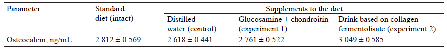
Osteocalcin in blood serum was quantified by enzyme-linked immunosorbent assay (ELISA) using a set of rat-specific reagents on an Immunochem 2100 analyzer (HTI, USA).
Statistical analysis was performed in STATISTICA 10. Statistical significance was determined by the Kruskal-Wallace H-test (P ≤ 0.05).
РЕЗУЛЬТАТЫ И ИХ ОБСУЖДЕНИЕ
First, we studied the physicochemical parameters of collagen fermentolysate. The molecular weight distribution of its fractions is shown in Fig. 1.
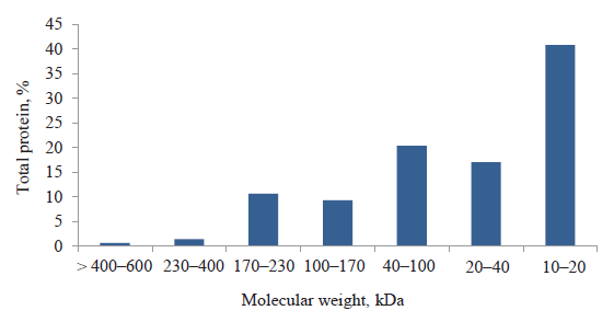
As can be seen in Fig. 1, about 41% of fractions weighed from 10 to 20 kDa. Peptides with such a molecular weight should be used as the basis of a functional beverage, since they ensure high bioavailability and good taste characteristics. The amino acid composition of collagen fermentolysate is shown in Fig. 2.
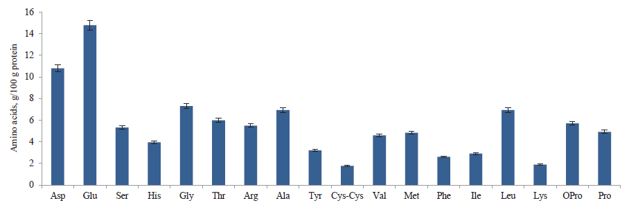
According Fig. 2, collagen fermentolysate contained relatively high contents of glutamic acid (14.8%), aspartic acid (10.8%), glycine (7.3%), alanine (6.9%), and proline (4.9%). These amino acids are known to stimulate cartilage and bone cells and restore joint tissues. Alanine is the main component of connective tissue, while proline and lysine are precursors of hydroxylysine and hydroxyproline, which are used by the body to form collagen, tendons, and heart muscle. We also found high contents of leucine and threonine. These essential amino acids are important for the biosynthesis of glycine and serine, which are responsible for the production of collagen, elastin, and muscle tissue.
Based on collagen fermentolysate, our functional product with an antiosteoporosis effect also contained dietary fiber, bioactive substances, as well as macro- and microelements.
The formulation was in line with the biomedical requirements for the quality, composition, and safety of functional products with corrective properties. To have a real physiological effect, the product should contain at least 50% of collagen fermentolysate. However, it should also have good consumer appeal. To neutralize the flavor of fermentolysate, pumpkin powder was used as dietary fiber. It has a pleasant taste and, at the same time, contains various carbohydrate components, including pectins, cellulose, fiber, calcium, magnesium, iron, B vitamins, vitamin PP, beta-carotene, and vitamin C.
Our formulation was primarily aimed at normalizing metabolic processes and preventing diseases of the musculoskeletal system. The component quantities met the physiological needs of adult humans.
Increased calcium intake is an integral part of osteoporosis prevention and treatment. To assimilate calcium, we added vitamin D3, as well as vitamins E and C with antioxidant properties. Products based on collagen hydrolysates in combination with vitamin C are more effective in stimulating collagen fibrils and proteoglycans in the cartilage matrix, thus improving joint mobility [19]. Oxidative stress is an important factor of aging that also contributes to osteoporosis. It induces bone resorption due to superoxide production by osteoclasts, which leads to bone degradation [5, 20, 21].
The dietary fiber included in the formulation is a prebiotic that ensures normal functioning of the gastrointestinal tract and has a beneficial effect on lipid and carbohydrate metabolism. In addition, indigestible oligosaccharides increase the absorption of various minerals, contributing to bone mineralization [22].
Of great importance are chondroprotectors – glucosamine sulfate and chondroitin sulfate. They have a positive effect on metabolic processes in cartilage tissue, slowing down degenerative changes in joints and the spine.
Our choice of ingredients was determined by two main objectives. Firstly, we aimed to formulate an efficient functional product with a high nutritional value. Secondly, we wanted this product to have good sensory characteristics. Both objectives could be achieved with pumpkin powder of the Gribovskaya variety. The pumpkin was peeled, cut into 5–8 mm pieces, blanched for 3–5 min, and placed on racks for 10–15 min to remove water. The pieces were then dried in a convection drying chamber in two stages – first, at 90 ± 5°С to a moisture content of 40–42% and then, at 60 ± 5°C to a moisture content of 3.0–5.0%. The dried pumpkin was crushed to a particle size of 0.2 mm. The resulting powder had a sweetish taste and yellowish color.
To determine an optimal ratio between collagen fermentolysate and dried pumpkin, in both functional and sensory terms, we carried out a sensory experiment. The panelists preferred the taste characteristics of a 80:20 ratio between protein hydrolysate and pumpkin powder. A higher content of hydrolysate gave the product a pronounced bitter taste, which was considered unacceptable.
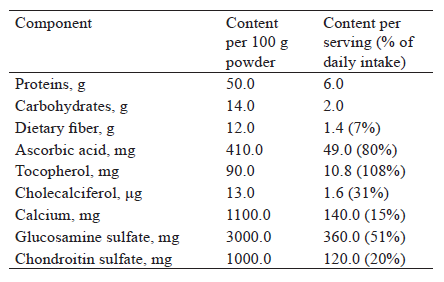
The amounts of functional ingredients had to meet the standard physiological needs without spoiling the consumer appeal. These ingredients included calcium lactate, glucosamine sulfate, chondroitin sulfate, ascorbic acid (vitamin C), tocopherol acetate (vitamin E), and cholecalciferol (vitamin D3). The formulated product is a dry powder for preparing a functional drink (Table 1).

Pre-mixtures based on the compatibility and fineness of ingredients were introduced into a drum-type mixer in two stages to ensure uniformity. The first premixture included fine ingredients in smaller quantities (vitamins D3, E, and C, chondroitin sulfate, and glucosamine sulfate). At the second stage, they were combined with the remaining components (protein hydrolysate, calcium lactate, dried pumpkin, and dietary fiber). A drink can be prepared by mixing 12 g of the concentrate with 100 mL of water. Three servings per day are needed to provide a good preventative effect.
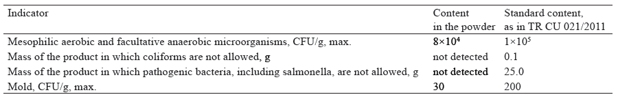
Next, we studied the sensory, physicochemical, microbiological, and toxicological indicators of the developed product. The nutritional value of the powdered product is shown in Table 2.
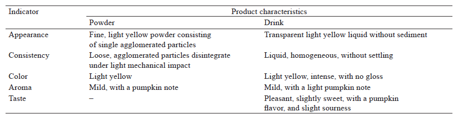
When formulating a functional product based on hydrolysates, it is important to crease a characteristic sensory profile. The sensory indicators of our functional product, both in powdered and ready-to-use form, are presented in Table 3.
The microbiological properties and contents of toxic substances (lead, arsenic, cadmium, and mercury) in the functional product were analyzed against the Technical Regulations of the Customs Union 021/2011 “On food safety” (Table 4 and Fig. 3).
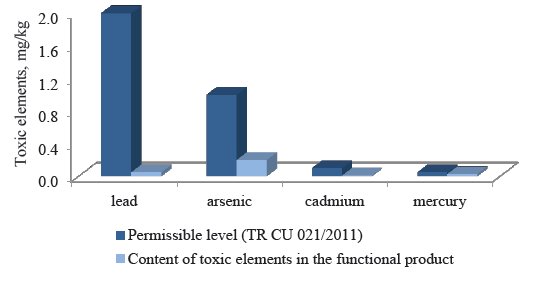
We found that the concentrations of lead and arsenic were significantly below the permissible values, and the contents of cadmium and mercury were within the norms established by TR CU 021/2011. This means that our functional product met the safety requirements of TR CU 021/2011.
Thus, we developed a powdered functional product based on collagen fermentolysate to prevent osteoporosis. Mixed with water, the drink can be used as an additional source of connective tissue protein, calcium, vitamins C, E, and D3, as well as dietary fiber and chondroprotectors.
Its effectiveness was confirmed in the experiment on laboratory animals with modelled osteoporosis (ovariectomized female rats).
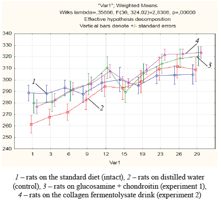
The weight of the intact animals was mostly stable throughout the experiment, with a slight increase from the 9th to the 15th day and from the 19th to the 26th day. The control animals, which received distilled water, gained weight during the entire experiment, especially from the 1st to the 12th day and from the 19th to the 26th day. Two groups of experimental animals, which received glucosamine + chondroitin and the drink based on collagen fermentolysate, also gained weight from the 1st to the 12th day and from the 15th to the 26th day. By the end of the experiment, the weight gain in the ovariectomized rats treated with distilled water, glucosamine + chondroitin, and the collagen fermentolysate drink was 16.0, 12.5, and 14.3%, respectively. The weight gain in the intact group was 5.3% (Fig. 4). Our data were consistent with the results of other studies [23]. Our findings were associated with a deficiency of estrogen that decreases the secretion of leptin (a hormone with anorexigenic effect) from adipose tissue, thus leading to hyperphagia.
Blood analysis showed a 29.6–60.5% increase in leukocytes in the ovariectomized animals, compared to the intact group. However, statistical significance was only registered in the group that received distilled water. A statistically significant (P < 0.05) increase of 46.5% in lymphocytes was observed in the rats treated with glucosamine + chondroitin (reference sample), compared to the intact animals. All the ovariectomized animals had an increased content of granulocytes (up to 86.4%), compared to the intact group, although we found significant variation between the experimental rats within the group. Yet, the increased concentrations of lymphocytes and granulocytes did not significantly affect their relative content.
The control rats treated with distilled water had a statistically insignificant increase in monocytes of 27.8% in relation to the intact group. This growth was more pronounced in both experimental groups (1 and 2), averaging 2.3 times (P < 0.05) in absolute terms and up to 47.8% (P < 0.05) in relative terms (Table 5).
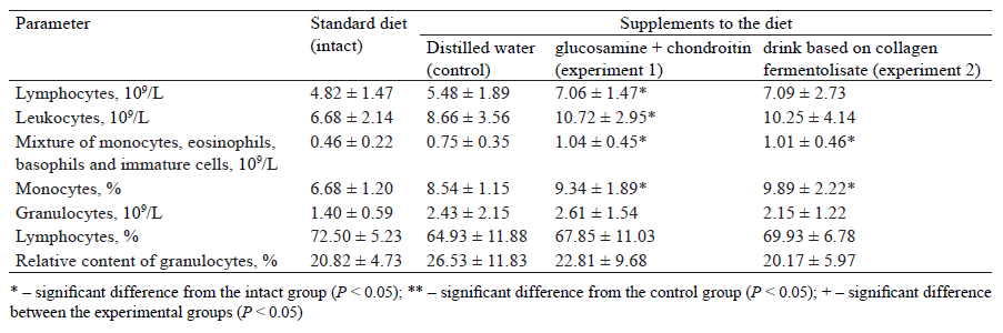
The cytometric analysis showed that both supplements to the diet (glucosamine + chondroitin and the functional drink) contributed to increased contents of leukocytes, granulocytes, lymphocytes, and monocytes, although to a different extent. This meant that they activated the blood immunity.
According to the biochemical blood analysis (Table 6), the control rats had a significant increase, compared to the intact rats, in alkaline phosphatase, phosphorus, and calcium by 36.0, 2.8, and 15.6%, respectively. They also had a significant decrease in magnesium by 15.3%. We know that increased concentrations of these parameters in the blood are among the diagnostic criteria for osteoporosis. However, the supplements of glucosamine + chondroitin and the functional drink decreased the activity of alkaline phosphatase to the intact level. This parameter in the experimental groups was 35.5% (P < 0.05) and 31.0% (P < 0.05) lower than in the control group. It might indicate a compensatory activation of the collagensynthetic function of osteoblastic cells in response to increased activity of osteoclasts.
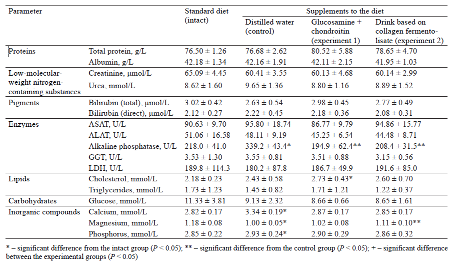
The results of the biochemical analysis were confirmed by the determination of osteocalcin.
Osteocalcin is the main non-collagenous protein of the bone matrix that is synthesized by osteoblasts. Its concentration in the blood reflects the metabolic activity of osteoblasts in bone tissue, since blood osteocalcin is synthesized, rather than released during bone resorption [24].
There were no statistically significant changes in osteocalcin concentrations between the groups (Table 7). However, we found its increase of 15.0% (statistically insignificant) in experimental group 2, which received the functional product based on collagen fermentolysate, compared to the control.
The contents of calcium, phosphorus, and magnesium in the bone tissue of the animals under study are shown in Table 8. As we can see, the control group of ovariectomized rats had decreased contents of calcium and magnesium (by 32.0 and 19.3%, respectively), compared to the intact group. They also had a significant increase in phosphorus levels (by 22.0%).
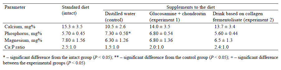
The experimental rats that received the functional drink had increased amounts of calcium and magnesium (by 25 and 3.0%, respectively), compared to the control group. Although we also found a 7.0% decrease in phosphorus, it was not statistically significant. The ratio between calcium and phosphorus in experimental group 2 was restored to 2.4:1.0 (2.5:1.0 in the intact group).
According to our daily examinations, the general condition of all the animals was satisfactory in terms of appearance, coat quality, and behavior. The experimental animals looked identical to the control group. Their coat was thick, tight, and glossy, with no signs of fur loss. They were physically strong and had no discharge from their natural orifices. Their limbs, motor functions, and behavioral reactions were normal. Their teeth were white, without plaque but with signs of abrasion. Their mucous membranes were pale, shiny, and smooth. The results of necropsy and macroscopic examination did not reveal any hypofunction or displacement in internal organs (lungs, liver, spleen, stomach, kidneys, and pancreas). Their pulmonary pleurae, as well as pericardial and abdominal layers, were thin, shiny, and smooth. The hearts and aortas were unchanged, and the vessels were moderately injected.
Some animals in the control and experimental groups had an enlarged uterus and mucus in the fallopian tubes. This might be associated with the involutional processes in their reproductive organs after surgery.
We weighed the animals’ internal organs and determined their percentage in relation to the body weight. The results revealed no significant differences from the physiological norms for the animals of this species and age group.
ВЫВОДЫ
We examined the quality characteristics of dried collagen fermentolysate obtained from low-value byproducts of the meat industry (pork legs and ears, 1:1). Collagen fermentolysate contained 41% of peptide fractions with a molecular weight of 10 to 20 kDa. It also had high contents of glutamic acid, aspartic acid, glycine, alanine, proline, leucine, and threonine. These amino acids stimulate cartilage and bone cells, restore joint tissue, and are responsible for the production of collagen, elastin, and muscle tissue.
We developed a technology for a functional product to prevent osteoporosis. Based on collagen fermentolysate, the formulation contained pumpkin powder, dietary fiber, calcium, chondroprotectors, and vitamins E, C, and D3. The 80:20 ratio between protein hydrolysate and pumpkin powder and the contents of calcium and vitamin D3 meeting 15 and 30% of the daily intake, respectively, ensured a high nutritional value, functional effects, and good sensory characteristics of the product. The product is a powdered drink designed to mix with water. The microbiological and toxicological analyses confirmed that the product complied with the requirements of TR CU 021/2011.
The experiments on laboratory animals showed that the formulated product had an osteoprotective effect on the ovariectomized female rats. We found that those rats which received the functional product had increased contents of calcium and magnesium in the bone tissue (by 25.0 and 3.0%, respectively) and a decreased content of phosphorus (by 7.0%), compared to the control group. In addition, their calcium to phosphorus ratio was restored to 2.4:1.0 and the concentration of osteocalcin in the blood serum increased by 15%.
Our study makes a theoretical contribution to the concept of safe bone homeostasis correction and proves that a functional drink based on connective tissue protein can be used to prevent postmenopausal osteoporosis associated with hypoestrogenism.
КОНФЛИКТ ИНТЕРЕСОВ
The authors declare that there is no conflict of interest.СПИСОК ЛИТЕРАТУРЫ
- Pisani P, Renna MD, Conversano F, Casciaro E, Di Paola M, Quarta E, et al. Major osteoporotic fragility fractures: Risk factor updates and societal impact. World Journal of Orthopedics. 2016;7(3):171–181. https://doi.org/10.5312/wjo.v7.i3.171.
- Srinivasan V, Martens MG. Hormone therapy in menopausal women with fibroids: is it safe?. Menopause. 2018;25(8):930–936. https://doi.org/10.1097/gme.0000000000001105.
- Muhammad A, Mada SB, Malami I, Forcados GE, Erukainure OL, Sani H, et al. Postmenopausal osteoporosis and breast cancer: The biochemical links and beneficial effects of functional foods. Biomedicine and Pharmacotherapy. 2018;107:571–582. https://doi.org/10.1016/j.biopha.2018.08.018.
- Rajput R, Wairkar S, Gaud R. Nutraceuticals for better management of osteoporosis: An overview. Journal of Functional Foods. 2018;47:480–490. https://doi.org/10.1016/j.jff.2018.06.013.
- Pinkerton JV, Aguirre FS, Blake J, Cosman F, Hodis H, Hoffstetter S, et al. The 2017 hormone therapy position statement of the North American Menopause Society. Menopause. 2017;24(7):728–753. https://doi.org/10.1097/gme.0000000000000921.
- Belyaev NG, Timchenko LD, Rzhepakovsky IV, Piskov SI, Lodygin AD, Gaponov VI, et al. Osteoprotective effect of bread enriched with protein, dietary fiber, calcium, iron and iodine in hypoestrogen-induced osteoporosis among rats. Problems of Nutrition. 2020;89(6):58–69. (In Russ.). https://doi.org/10.24411/0042-8833-2020-10079.
- Liu J, Liu J, Liu L, Zhang G, Zhou A, Peng X. The gut microbiota alteration and the key bacteria in Astragalus polysaccharides (APS)-improved osteoporosis. Food Research International. 2020;138. https://doi.org/10.1016/j.foodres.2020.109811.
- Honvo G, Lengelé L, Charles A, Reginster J-Y, Bruyère O. Role of collagen derivatives in osteoarthritis and cartilage repair: A systematic scoping review with evidence mapping. Rheumatology and Therapy. 2020;7:703–740. https://doi.org/10.1007/s40744-020-00240-5.
- Kakimov A, Suychinov A, Mayorov A, Yessimbekov Z, Okuskhanova E, Kuderinova N, et al. Meat-bone paste as an ingredient for meat batter, effect on physicochemical properties and amino acid composition. Pakistan Journal of Nutrition. 2017;16(10):797–804. https://doi.org/10.3923/pjn.2017.797.804.
- Nasri M. Protein hydrolysates and biopeptides: Production, biological activity, and application in food and health benefits. A review. Advances in Food and Nutrition Research. 2018;81:109–159. https://doi.org/10.1016/bs.afnr.2016.10.003.
- Kiewiet MBG, Faas MM, de Vos P. Immunomodulatory protein hydrolysates and their application. Nutrients. 2018;10(7). https://doi.org/10.3390/nu10070904.
- López-Pedrouso M, Borrajo P, Pateiro M, Lorenzo JM, Franco D. Antioxidant activity and peptidomic analysis of porcine liver hydrolysates using alcalase, bromelain, flavourzyme and papain enzymes. Food Research International. 2020;137. https://doi.org/10.1016/j.foodres.2020.109389.
- Inoue N, Sugihara F, Wang X. Ingestion of bioactive collagen hydrolysates enhance facial skin moisture and elasticity and reduce facial ageing signs in a randomised double-blind placebo-controlled clinical study. Journal of the Science of Food and Agriculture. 2016;96(12):4077–4081. https://doi.org/10.1002/jsfa.7606.
- Shi P, Liu M, Fan F, Chen H, Yu C, Lu W, et al. Identification and mechanism of peptides with activity promoting osteoblast proliferation from bovine lactoferrin. Food Bioscience. 2018;22:19–25. https://doi.org/10.1016/j.fbio.2017.12.011.
- Hong H, Fan H, Chalamaiah M, Wu J. Preparation of low-molecular-weight, collagen hydrolysates (peptides): Current progress, challenges, and future perspectives. Food Chemistry. 2019;301. https://doi.org/10.1016/j.foodchem.2019.125222.
- Yunusov EhSh, Ponomarev VYa, Morozova SA, Ezhkova GO. Izuchenie gidroliza kollagensoderzhashchego syrʹya proteoliticheskimi fermentami [A study of hydrolysis of collagen-containing materials with proteolytic enzymes]. Bulletin of the Technological University. 2016;19(24):168–170. (In Russ.).
- Aslanova MA, Dydykin AS, Soldatova NE. Preparation of protein hydrolyzate from raw materials of animal origin for the enrichment of products. Food Industry. 2018;(2):16–18. (In Russ.).
- Chernukha IM, Kotenkova EA, Vasilevskaya ER, Ivankin AN, Lisitsyn AB, Fedulova LV. The study of biological effects of different geographical origin goji berries in rats with alimentary hypercholesterolemia. Problems of Nutrition. 2020;89(1):37–45. (In Russ.). https://doi.org/10.24411/0042-8833-2020-10004.
- Nikolaeva TI, Shekhovtsov PV. Hydrolysates of collagen concerning prevention and healing joint diseases. Fundamental research. 2014;(12–3):524–528. (In Russ.).
- Marchenkova LA, Fesyun AD, Gerasimenko MYu, Makarova EV. The effect of administration of dietary supplement with calcium and vitamins D3 and B6 on calcium homeostasis and falls incidence in patients with high fracture risk undergoing medical rehabilitation. Problems of Nutrition. 2020;89(5):89–100. (In Russ.). https://doi.org/10.24411/0042-8833-2020-10069.
- Heaney RP, Bone as the calcium nutrient reserve. In: Weaver CM, Heaney RP, editors. Calcium in human health. Totowa, New Jersey: Humana Press; 2006. pp. 7–12. https://doi.org/10.1007/978-1-59259-961-5_2.
- Scholz-Ahrens KE, Schaafsma G, Van den Heuvel EGHM, Schrezenmeir J. Effects of prebiotics on mineral metabolism. The American Journal of Clinical Nutrition. 2001;73(2):459S–464S. https://doi.org/10.1093/ajcn/73.2.459s.
- Puzikov AM. Osteoporosis correction in experiment. Experimental and Clinical Gastroenterology Journal. 2014;102(2):25–31. (In Russ.).
- Klimova ZhA, Zaft AA, Zaft VB. Modern laboratory diagnosis of osteoporosis. International Journal of Endocrinology. 2014;63(7):75–84. (In Russ.).
