

Аннотация
Introduction. This study aimed to evaluate the antioxidant and anti-diabetic activity of aqueous and hydroalcoholic extracts of pomegranate (Punica granatum L.) leaves in vitro, as well as to determine the content of polyphenols, flavonoids, and flavonols.Study objects and methods. The antioxidant activity was determined by the DPPH test using the free radical 1,1-diphenyl-2-picrylhydrazyle and the FRAP method, as well as by measuring total antioxidant capacity and the hydrogen peroxide scavenging activity.
Results and discussion. The content of total polyphenols varied between 4.43 ± 0.3 and 12.66 ± 1.6 mg EAG/g. The highest content of flavonoids was observed in the hydroalcoholic extract of P. granatum leaves (P < 0.05). The flavonol contents in the hydroalcoholic and aqueous extracts were 7.68 ± 0.6 and 9.20 ± 2.8 mg EQ/g, respectively. The IC50 of the antioxidant potential of the hydroalcoholic and aqueous extracts was 32.4 ± 1.109 and 35.12 ± 4.107 mg/mL, respectively. According to the DPPH test, the aqueous extract was the least active (IC50 = 14.15 ± 1.513 mg/mL). The highest percentage of hydrogen peroxide trapping was found in the aqueous extract (45.97 ± 6.608 %). The inhibition of α-amylase showed an IC50 of between 9.804 ± 0.67 and 19.011 ± 9.82 mg/mL in the aqueous and hydroalcoholic extracts, respectively. The inhibition of glucose uptake by yeast recorded a high inhibitory capacity at 50 mg/mL of glucose.
Conclusion. We found that the antioxidant and anti-diabetic activity of P. granatum leaves extracts was due to the presence of bioactive compounds such as flavonoids, which is why they are effective in preventing diabetes and its complications.
Ключевые слова
Punica granatum L., plant extracts, antioxidant activity, anti-diabetic activity, flavonoidsВВЕДЕНИЕ
The pomegranate (Punica granatum L.) is a shrub that belongs to the Lythraceae family. It is between 5 and 10 m tall and is characterized by deciduous fruiting leaves. The pomegranate is used to prevent cancer, cardiovascular disease, diabetes, dental conditions, and erectile dysfunction, as well as against ultra violet radiation. Pomegranate leaf extracts contain high total phenols, tannins, and triterpenoids [1].
Numerous studies have demonstrated the in vitro antioxidant activity and polyphenol content of pomegranate. According to Amjad et al., the antioxidant activity of pomegranate leaves is directly related to the presence of phenolic compounds and antioxidant components which act as hydrogen donors, contributing to the concentration of total phenols [2]. These authors demonstrated that pomegranate n-butanol, ethyl acetate, hydroethanol, and aqueous leaf extracts contained ellagic acid, an efficient free radical scavenger [2].
Vinodhini et al. reported that the aqueous extract of pomegranate leaves had the greatest antioxidant activity and contained significant levels of total phenols and flavonoids [3]. The leaf extracts showed antioxidant activity in vivo by protecting yeast cells against oxidative stressing agent H2O2. The authors found pomegranate a good source of natural compounds with health benefits, which makes it possible to use it in diets to reduce oxidative stress.
In the study by Bekir et al., the methanolic extract of pomegranate leaves displayed high antioxidant, antiinflammatory, anti-cholinesterase, and antiproliferative activities [4]. These results showed that pomegranate leaves could be a potential source of active molecules intended for applications in pharmaceutical industry.
The aim of the study was to evaluate the antioxidant and anti-diabetic activity of aqueous and hydroalcoholic extracts of pomegranate leaves in vitro.
ОБЪЕКТЫ И МЕТОДЫ ИССЛЕДОВАНИЯ
The pomegranate (Punica granatum L.) leaves were collected in September 2017 in Chlef, Algeria. The collected samples were dried at room temperature away from sunlight and then powdered using an electric mortar.
Preparation of aqueous extract. The aqueous extract of pomegranate leaves was prepared according to the method described by Diallo et al., with some modifications [5]. 15 g of powdered leaves in 150 mL of boiling water was heated for 15 min and filtered through filter paper. The filtrate was placed in an oven at 40°C until obtaining a dry extract and stored at 4°C.
Preparation of hydroalcoholic extract. The hydroalcoholic extract of pomegranate leaves was prepared by maceration of 15 g of powdered leaves in 100 mL of a hydroalcoholic solution (70%) at room temperature away from light, with maximum agitation for 72 h [6]. Then the mixture was filtered through filter paper. The filtrates were placed in an oven at 40°C. The dry extract was stored in a refrigerator at 4°C.
Total polyphenols were determined spectrophotometrically following the Folin-Cioclateu method [7]. For this, 0.2 mL of each leaf extract was mixed in a test tube with 1.0 mL of Folin-Cioclateu reagent and 0.8 mL of a 7.5% sodium carbonate solution (Na2CO3). After incubation in the shade and at room temperature for 30 min, absorbance was measured at 760 nm. The results were expressed in milligram equivalent of gallic acid per gram of extract (mg EAG/g extract) from a calibration curve prepared using gallic acid as a standard.
Flavonoid levels were measured using the method described by Mbaebie et al. [8]. For this, 1.0 mL of each extract was added to 1.0 mL of a 2% ethanol solution of aluminum chloride (AlCl3) and then incubated for an hour at room temperature. Absorbance was measured by a UV-visible spectrophotometer at 420 nm. The concentrations of flavonoids in the extracts were calculated from the calibration curve and expressed in milligram equivalent of quercetin per gram of extract (mg EQ/g extracted).
Flavonols were determined according to the method described by Kosalec et al. [9]. For this, 0.3 mL of the extract was mixed with 0.3 mL of aluminum chloride (AlCl3) and 0.45 mL of sodium acetate. The mixture was vigorously stirred and then incubated for 40 min. Absorbance was measured at 440 nm. The quantification of flavonols was based on a calibration curve made by quercetin. The content of flavonols was expressed in milligram equivalent of quercetin per gram of extract (mg EQ/g).
Total antioxidant capacity. Determination of total antioxidant capacity is a technique based on the reduction of molybdate Mo (VI) to molybdenum Mo (V) in the presence of an antioxidant with the formation of a green complex (phosphate/Mo (V)) at acidic pH [10]. The phosphomolybdate reagent was prepared from a mixture of 0.6 M sulfuric acid (H2SO4), 28 mM sodium phosphate (Na3PO4), and 4 mM ammonium molybdate ((NH4)6Mo7O24 • 4H2O). 1.0 mL of this reagent was added to 100 μL of each extract with concentrations of 10, 25, 50 and 100 mg/mL. The tubes were incubated at 95°C for 90 min. After cooling, absorbance was measured at 695 nm. Total antioxidant capacity was expressed in milligrams of ascorbic acid equivalent per gram of extract (mg Eq AA/g extract) from a calibration curve of ascorbic acid.
Ferric Reducing Antioxidant Power (FRAP). The FRAP method involves measuring the ability of a sample to reduce the tripyridyltriazine ferric complex to tripyridyltriazine at a low pH. This ferrous tripyridyltriazine complex has an intense blue color measured by a spectrophotometer at 593 nm [11]. The FRAP reagent was prepared by mixing a 300 mM sodium acetate buffer (pH 3.6), a solution of 10 mM TPTZ in 40 mM HCl and 20 mM FeCl3 in a ratio of 10:1:1 (v/v/v). 200 μL of each extract (10, 25, 50 and 100 mg/mL) was added to 3 mL of the FRAP reagent. After incubation in the dark at 37°C for 30 min, absorbance was measured at 593 nm against the blank [11].
Hydrogen peroxide scavenging activity. The scavenging capacity of hydrogen peroxide is based on the reduction of the H2O2 concentration by scavenger compounds, the absorbance value of the latter at 230 nm also reduces [12]. A 40 mM hydrogen peroxide solution was prepared in a 50 mM phosphate buffer (pH 7.4). 4.0 mL of each extract with a concentration of 10 mg/mL was mixed with 0.6 mL of the H2O2 solution. After 10 min incubation, absorbance was measured at 230 nm. Ascorbic acid was used as a positive control [13]. The percent inhibition was calculated using the following equation:
where A is absorbance of the control and experimental samples.
DPPH test. To p repare a 0.004% solution of DPPH, 250 μL of extracts at concentrations of 10, 25, 50, and 100 mg/mL or standard (ascorbic acid) was added to 1 mL of the DPPH solution. After incubation in the dark at room temperature for 30 min, absorbance was measured at 517 nm against a blank sample that contained pure methanol [14]. The antioxidant activity evaluated with the DPPH method was expressed in percentage according to the following formula:
where A is absorbance of the control and experimental samples.
Inhibition test of α-amylase enzymatic activity. The inhibition test of α-amylase enzymatic activity followed the method of Daksha et al. [15]. For t his, t wo solutions had been prepared, namely a 1% starch stock solution and a 1% amylase solution in a 0.1 M phosphate buffer at pH 7.2, both solutions preserved at 4°C. The reaction mixture contained 2.0 mL of a phosphate buffer, 1.0 mL of each extracts (aqueous and hydroalcoholic) at concentrations of 10, 25, 50, and 100 mg/mL, 1 mL of amylase, and 1 mL of starch. The mixture was incubated for an hour. The enzymatic reaction was stopped by the addition of 0.1 mL of the iodide indicator. All experiments were performed in triplicate. Absorbance was measured at 565 nm.
The inhibitory activity of each extract was calculated according to the following formula:
To determine effects of the extracts on glucose uptake by yeast, we prepared yeast cells according to the method described in [16]. 1 g of commercial baker’s yeast was washed by centrifugation (4200 rpm, 5 min) in 5 mL of distilled water until the supernatant liquid was clear. Then a 10% suspension (v/v) was prepared in distilled water. Different concentrations of plant extracts (10 to 100 mg/mL) were added to 1 mL of glucose solution (10, 25 and 50 mg/mL) and incubated together for 15 min at 37°C. Then, 100 μL of the yeast suspension was added, followed by a vortex and a new incubation at 37°C for 60 min. After one hour, the tubes were centrifuged (2500 rpm, 5 min) and glucose was estimated in the supernatant by the iodine reagent [17]. Metformin was taken as a standard antidiabetic drug. Absorbance was measured at 540 nm and all experiments were performed in triplicate. The percentage increase in glucose uptake by yeast cells was calculated using the following formula [18]:
The data presented in our study were analyzed using XL Stat Pro 7.5 statistical software. The experiments were performed in triplicate. The results were presented as mean values and a standard deviation. ANOVA test was conducted to determine any significance differences. P < 0.05 was considered as statistically significant.
РЕЗУЛЬТАТЫ И ИХ ОБСУЖДЕНИЕ
Table 1 demonstrates total phenolic, flavonoid and flavonol contents of the pomegranate (Punica granatum L.) extracts. The hydroalcoholic extract showed a significantly (P < 0.05) higher content of total phenolic compounds compared to the aqueous extract, with values of 12.66 ± 0.10 and 4.43 ± 0.01 mg EAG/g extract, respectively (Table 1). These results were not consistent with those found by Sinha et al., namely 9.85 ± 0.82 and 14.78 ± 2.10 mg EAG/g extract for the pomegranate aqueous and hydroalcoholic extracts, respectively [19].

The hydroalcoholic extract showed a significantly (P < 0.05) higher content of flavonoids than the aqueous extract (24.78 ± 1.59 and 8.76 ± 0.90 mg EQ/g, respectively). These results were closer to those reported by [19], namely 12.7 ± 0.23 and 26.08 ± 1.24 mg EQ/g for the aqueous and methanolic extracts, respectively. According to quantitative analyses, pomegranate leaves contained a higher amount of flavonoids compared to phenolic compounds. These results were confirmed by [19], where pomegranate leaf extracts showed a lower content of total polyphenols and a higher content of flavonoids compared to pomegranate bark, flower, and seed extracts.
Our results indicated that the aqueous extract was richer in flavonols compared to the hydro-alcoholic extract; with contents of 9.20 ± 2.80 and 7.68 ± 0.60 mg EQ/g of extract, respectively (Table 1). The statistical analyses did not show any significant difference between the two extracts (P > 0.05).
Table 2 shows the antioxidant capacity of the pomegranate extracts. The aqueous extract of pomegranate leaves had a significantly higher (P < 0.05) total antioxidant capacity with an IC50 value of 12.404 ± 0.136 mg/mL, while the hydroalcoholic extract showed a significantly lower (P > 0.05) antioxidant capacity with an IC50 of 18.719 ± 1.001 mg/mL.

In a study of three local varieties of Piper betle leaves by Dasgupta et al., the Kauri variety showed the highest total antioxidant capacity expressed in milligrams of ascorbic acid equivalent per milligram of extract [20].
According to the FRAP test results, the antioxidant potential of iron was almost the same for both hydroalcoholic and aqueous extracts, with IC50 of 32.4 ± 1.109 and 35.12 ± 4.107 mg/mL, respectively (Table 2).
While there were no significant differences (P > 0.05) between the hydroalcoholic and aqueous extracts, there was a significant difference (P < 0.05) between the extracts and ascorbic acid, which showed a reducing power with an IC50 of 55.531 ± 1.133 mg/mL.
These results were not consistent with those recorded by [19], namely IC50 of 348.68 ± 24.69 and 293.63 ± 15.29 mg/mL for the aqueous and methanolic extracts of pomegranate leaves, respectively.
The percentage of hydrogen peroxide scavenging activity of the hydroalcoholic and aqueous extracts was 43.57 ± 10.145% and 45.97 ± 6.608%, respectively. There was no significant difference between the extracts (P > 0.05) (Table 2).
Compared to the extracts, ascorbic acid showed a significantly higher (P < 0.05) percentage, namely 85.663 ± 5.024%.
According to the DPPH test results, the hydroalcoholic extract was significantly the most potent extract (P < 0.05) w ith a n IC50 of 9.40 ± 1.586 mg/mL, followed by the aqueous extract with an IC50 of 14.15 ± 1.513 mg/mL (Table 2).
Compared to the extracts, the standard antioxidant (ascorbic acid) showed a significantly higher (P < 0.05) antioxidant activity, with an IC50 of 2.27 ± 0.012 mg/mL (Table 2).
These results were in agreement with the data reported by Bekiretal, where the methanolic extract of pomegranate leaves showed a greater antioxidant activity than the ethanolic extract, with an IC50 of 5.62 ± 0.23 mg/L and 9.25 ± 0.72 mg/L, respectively [4]. The study also revealed comparable antioxidant activity between the methanolic extract and quercetin (2.86 ± 0.09 mg/L). The dichloromethane extract showed lower antioxidant activity (IC50 = 71.57 ± 3.65mg/L). However, the extract obtained with hexane had the lowest DPPH activity with an IC50 value of 263.44 ± 12.72 mg/L.
According to Fig. 1, the aqueous extract showed an α- amylase inhibitory concentration of 9.804 ± 0.67 mg/mL. This value was significantly lower (P < 0.05) than that for acarbose and hydroalcoholic extracts, with IC50 values of 17.179 ± 4.26 and 19.011 ± 9.82 mg/mL, respectively. On the other hand, there was no significant difference between the IC50 of acarbose and the IC50 of the hydroalcoholic extract (P > 0.05).
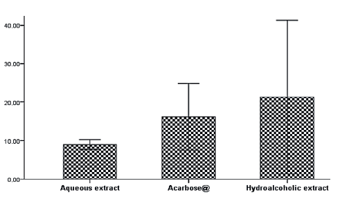
These inhibition results were not in agreement with those found by Kam et al., who recorded IC50 inhibitory concentrations of 0.19 and 0.65 mg/mL for aqueous and alcoholic extracts of pomegranate, respectively [21].
This inhibitory power can be explained by the fact that the hydroalcoholic and aqueous extracts have compounds that bear functional groups close to those of the substrate (starch), which occupies the active site of the enzyme.
Figure 2 demonstrates the inhibition of glucose uptake by yeast. At a concentration of 10 mg/mL of glucose, metformin showed a significant difference from the extracts (P < 0.05), with an IC50 of 5.442 ± 0.047 mg/mL. However, we found no significant difference between the hydroalcoholic and aqueous extracts (P > 0.05), with IC50 values of 7.267 ± 0.644 and 6.975 ± 0.394 mg/mL, respectively.
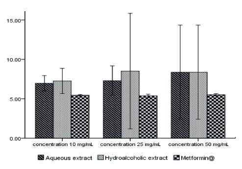
At a concentration of 25 mg/mL of glucose, there was no significant difference (P > 0.05) between the aqueous extract, metformin, and hydro-alcoholic extract, with IC50 values of 7.297 ± 0.76, 5.353 ± 0.11, and 8.509 ± 2.94 mg/mL, respectively (Fig. 2).
At a concentration of 50 mg/mL of glucose, there was a significant difference between metformin and the extracts (P < 0.05) and no significant difference (P > 0.05) between the extracts. The IC50 values of metformin, aqueous and hydroalcoholic extracts were 5.499 ± 0.073, 8.379 ± 2.4, and 8.937 ± 2.892 mg/mL, respectively (Fig. 2).
Based on these results, metformin showed a higher inhibition capacity than the aqueous and hydroalcoholic extracts.
According to the results, the antioxidant property varied according to the extraction solvent. The antioxidant properties of plant extracts can be explained by various factors: the presence of natural ascorbic acid (vitamin C), α-tocopherol (vitamin E), β-carotene (a precursor of vitamin A), flavonoids, and other phenolic compounds [22, 23].
These phenolic compounds are capable of acting as antioxidants that can neutralize free radicals by donating an electron or a hydrogen atom [24, 25].
The antioxidant capacity of phenolic compounds is also attributed to their ability to chelate ionic metals involved in the production of free radicals. For example, when attaching a ligand (phenolic compound) to Fe+3 in the FRAP test, polyphenols can reduce iron to Fe+2 [26].
Antioxidants act as “sensors” of free radicals, fighting against radical oxidation. Antioxidants of phenolic type react according to a mechanism proposed by Sherwin in 1976: an antioxidant formally yields a hydrogen radical, which may be an electron transfer followed, more or less rapidly, by a proton transfer [27].
Polyphenolic compounds are increasingly being used in therapeutics [28]. Many studies suggest that polyphenols participate in the prevention of cardiovascular diseases. They inhibit the oxidation of low density lipoproteins and platelet aggregation involved in the phenomenon of thrombosis that can lead to occlusion of the arteries [29]. These compounds show antioxidant activities: they have anti-inflammatory, antiatherogenic, antithrombotic, analgesic, antibacterial, and antiviral effects and can act as anticarcinogens, antiallergens, or vasodilators [30, 31].
Flavonoids also perform many biological functions that are attributed in part to their antioxidant properties. These compounds not only inhibit free radicals, but also neutralize oxidative enzymes and chelate metal ions responsible for the production of reactive oxygen species [32].
As for tannins, they are defined as sources of plant origin because they can precipitate proteins, inhibit digestive enzymes, and decrease the use of vitamins and minerals. On the other hand, tannins are also considered as “health promoting” components in plant-derived foods and beverages. For example, tannins have been reported to have anti-carcinogenic and antimutagenic potential, as well as antimicrobial properties.
The antioxidant activity of pomegranate leaves is due to their richness in phenolic compounds (tannins, flavones, glucosides). In fact, the work by Kang et al. suggested that polar polyphenolic molecules present in the plant’s extract contributed to the increase in antiradical activity [33].
As for anti-diabetic activity, Patel et al. reported that pomegranate extract regulates post-ponderal glucose by its inhibitory effect on α-amylase [34].
Flavonoids have a high nutritional value because they are part of our usual diet, which could be explained by their rapid metabolism, elimination, and relatively low bioavailability [35].
The reaction mechanisms of α-amylase enzyme inhibition remain unclear. However, flavonoids in foods can interact with starch and react with nitrous acid derived from the oral cavity in the stomach before being transported to the intestine [36]. This review mainly deals with: (a) the inhibition of α-amylase activity by flavonoids, suggesting the mechanisms of inhibition, and (b) the suppression of starch digestion by flavonoids by forming starch-flavonoid complexes through hydrophobic interactions.
The inhibition potential for flavonoids and tannins is correlated with the number of hydroxyl groups in their B cycles. These compounds inhibit α-amylase by forming hydrogen bonds between its hydroxyl groups and the residues of the active site of this enzyme. Flavonoids or flavonoid-rich foods can reduce the risk of diabetes by modulating glucose uptake and insulin secretion [37].
The transport of glucose through the yeast cell membrane occurs by facilitated diffusion, a passive mechanism without energy input. Glucose transport is continued if intracellular glucose is effectively reduced or used [38].
Scientific evidence shows that apical or luminal GLUT 2, facilitating the intestinal transport of glucose, is the major route of glucose uptake and thus an attractive target for some plant-based inhibitory agents [39].
Calystegine, a compound found in the pomegranate, exerts an antidiabetic effect by acting on the absorption of glucose by a competitive mechanism because of their structural analogy with glucose [40].
ВЫВОДЫ
Our study demonstrated that pomegranate (Punica granatum L.) leaf extracts are rich in phenolic compounds which play a very important role in the scavenging of free radicals, it makes a significant contribution to the justification of the antioxidant and anti-diabetic activity. It gives the extracts a power to protect the body against stress and manifestations linked to diabetes. The hydroalcoholic leaves extract was effective in preventing diabetes due to its high flavonoid. Therefore, there is a need for further in vivo studies to better understand the mechanism of their action.КОНФЛИКТ ИНТЕРЕСОВ
The authors declare that there is no conflict of interests.СПИСОК ЛИТЕРАТУРЫ
- Sreedevi P, Vijayalakshmi K, Venkateswari R. Phytochemical evaluation of Punica granatum L. leaf extract. International Journal of Current Pharmaceutical Research. 2017;9(4):14–18. DOI: https://doi.org/10.22159/ijcpr.2017v9i4.1159.
- Amjad L, Shafighi M. Antioxidant activity of leaf different extracts in Punica granatum. International Journal of Biological and Medical Research. 2012;3(3):2065–2067.
- Vinodhini S, Shri Preethi M, Nusrath Fathima N, Kushwaha SS, Devi Rajeswari V. Antioxidant and free radical scavenging capacity extensively used medicinal plant of Punica granatum. Asian Journal of Pharmaceutical and Clinical Research. 2016;9(6):140–146. DOI: https://doi.org/10.22159/ajpcr.2016.v9i6.13941.
- Bekir J, Mars M, Souchard JP, Bouajila J. Assessment of antioxidant, anti-inflammatory, anti-cholinesterase and cytotoxic activities of pomegranate (Punica granatum) leaves. Food and Chemical Toxicology. 2013;55:470–475. DOI: https://doi.org/10.1016/j.fct.2013.01.036.
- Diallo D, Sanogo R, Yasambou H, Traoré A, Coulibaly K, Maïga A. Étude des constituants des feuilles de Ziziphus mauritiana Lam. (Rhamnaceae), utilisées traditionnellement dans le traitement du diabète au Mali. Comptes Rendus Chimie. 2004;7(10–11):1073–1080. DOI: https://doi.org/10.1016/j.crci.2003.12.035.
- Cheurfa M, Allem R. Evaluation of antioxidant activity of different extracts of Aloysia triphylla leaves (L’Herit.) from Algeria in vitro. Phytotherapie. 2016;14(3):181–187. DOI: https://doi.org/10.1007/s10298-015-0969-4.
- Singleton VL, Orthofer R, Lamuela-Raventós RM. Analysis of total phenols and other oxidation substrates and antioxidants by means of folin-ciocalteu reagent. Methods in Enzymology. 1999;299:152–178. DOI: https://doi.org/10.1016/S0076-6879(99)99017-1.
- Mbaebie BO, Edeoga HO, Afolayan AJ. Phytochemical analysis and antioxidants activities of aqueous stem bark extract of Schotia latifolia Jacq. Asian Pacific Journal of Tropical Biomedicine. 2012;2(2):118–24. DOI: https://doi.org/10.1016/S2221-1691(11)60204-9.
- Kosalec I, Pepeljnjak S, Bakmaz M, Vladimir-Knezević S. Flavonoid analysis and antimicrobial activity of commercially available propolis products. Acta Pharmaceutica. 2005;55(4):423–230.
- Prieto P, Pineda M, Aguilar M. Spectrophotometric quantitation of antioxidant capacity through the formation of a phosphomolybdenum complex: Specific application to the determination of vitamin E. Analytical Biochemistry. 1999;269(2):337–341. DOI: https://doi.org/10.1006/abio.1999.4019.
- Benzie IFF, Strain JJ. The ferric reducing ability of plasma (FRAP) as a measure of “antioxidant power”: The FRAP assay. Analytical Biochemistry. 1996;239(1):70–76. DOI: https://doi.org/10.1006/abio.1996.0292.
- Magalhaes LM, Segundo MA, Reis S, Lima J. Methodological aspects about in vitro evaluation of antioxidant properties. Analytica Chimica Acta. 2008;613(1):1–19. DOI: https://doi.org/10.1016/j.aca.2008.02.047.
- Nagulendran K, Velavan S, Mahesh R, Begum VH. In vitro antioxidant activity and total polyphenolic content of Cyperus rotundus rhizomes. Journal of Chemistry. 2007;4(3):440–449. DOI: https://doi.org/10.1155/2007/903496.
- Brand-Williams W, Cuvelier ME, Berset C. Use of a free-radical method to evaluate antioxidant activity. LWT – Food Science and Technology. 1995;28(1):25–30.
- Daksha G, Subraya Chandrashekar K, and Pai G. In vitro antidiabetic activity of pentacyclic tritrpenoids and fatty acid esters from Bauhinia Purpurea. International Journal of Pharmacology and Pharmaceutical Technology. 2013;2(1):25–28.
- Nair SS, Kavrekar V, Mishra A. Evaluation of in vitro anti diabetic activity of selected plant extracts. International Journal of Pharmaceutical Science Invention. 2013;2(4):12–19.
- Sinha SK, Ahmad I, Gayathri M. Antidiabetic effect of ethanol extract of Syzygium jambolanum seed (in-vitro). International Journal of Drug Development and Research. 2013;5(3):187–191.
- Ukwubile CA, Odugu JA. Evaluation of antibacterial and in vitro antidiabetic activities of Phyllanthus amarus Linn. (Phyllanthaceae) leaf ethanol extract. Journal of Bacteriology and Mycology: Open Access. 2018;6(4):254–256. DOI: https://doi.org/10.15406/jbmoa.2018.06.00214.
- Elfalleh W, Hannachi H, Tlili N, Yahia Y, Nasri N, Ferchichi A. Total phenolic contents and antioxidant activities of pomegranate peel, seed, leaf and flower. Journal of Medicinal Plants Research. 2012;6(32):4724–4730. DOI: https://doi.org/10.5897/JMPR11.995.
- Dasgupta N, De B. Antioxidant activity of Piper betle L. leaf extract in vitro. Food Chemistry. 2004;88(2):219–224. DOI: https://doi.org/10.1016/j.foodchem.2004.01.036.
- Kam A, Li KM, Razmovski-Naumovski V, Nammi S, Shi J, Chan K, et al. A comparative study on the inhibitory effects of different parts and chemical constituents of pomegranate on α-amylase and α-glucosidase. Phytotherapy Research. 2013;27(11):1614–1620. DOI: https://doi.org/10.1002/ptr.4913.
- Oleynikov VV. Antioxidant and antimicrobial properties of oregano extract (Origani vulgaris herba L.). Foods and Raw Materials. 2020;8(1):84–90. DOI: https://doi.org/10.21603/2308-4057-2020-1-84-90.
- Pennington JAT, Fisher RA. Classification of fruits and vegetables. Journal of Food Composition and Analysis. 2009;22:S23–S31. DOI: https://doi.org/10.1016/j.jfca.2008.11.012.
- Laughton MJ, Halliwell B, Evans PJ, Hoult JRS. Antioxidant and pro-oxidant actions of the plant phenolics quercetin, gossypol and myricetin: Effects on lipid-peroxidation, hydroxyl radical generation and bleomycin-dependent damage to DNA. Biochemical Pharmacology. 1989;38(17):2859–2865. DOI: https://doi.org/10.1016/0006-2952(89)90442-5.
- Apak R, Guclu K, Demirata B, Ozyurek M, Celik SE, Bektasoglu B, et al. Comparative evaluation of various total antioxidant capacity assays applied to phenolic compounds with the CUPRAC assay. Molecules. 2007;12(7):1496–1547. DOI: https://doi.org/10.3390/12071496.
- Perron NR, Brumaghim JL. A review of the antioxidant mechanisms of polyphenol compounds related to iron binding. Cell Biochemistry and Biophysics. 2009;53(2):75–100. DOI: https://doi.org/10.1007/s12013-009-9043-x.
- Nkhili E. Polyphénols de l’Alimentation: Extraction, Interactions avec les ions du Fer et du Cuivre, Oxydation et Pouvoir antioxidant [Polyphenols from the feed: Extraction, Interactions with iron and copper ions, oxidation and antioxidant power]. Dr. eng. sci. diss. Montpellier: Université D’Avignon; 2009. 378 p. (In French).
- Crozier A, Del Rio D, Clifford MN. Bioavailability of dietary flavonoids and phenolic compounds. Molecular Aspects of Medicine. 2010;31(6):446–467. DOI: https://doi.org/10.1016/j.mam.2010.09.007.
- Manach C, Mazur A, Scalbert A. Polyphenols and prevention of cardiovascular diseases. Current Opinion in Lipidology. 2005;16(1):77–84. DOI: https://doi.org/10.1097/00041433-200502000-00013.
- Han XZ, Shen T, Lou HX. Dietary polyphenols and their biological significance. International Journal of Molecular Sciences. 2007;8(9):950–988. DOI: https://doi.org/10.3390/i8090950.
- Hodgson JM, Croft KD. Tea flavonoids and cardiovascular health. Molecular Aspects of Medicine. 2010;31(6):495–502. DOI: https://doi.org/10.1016/j.mam.2010.09.004.
- Cotelle N. Role of flavonoids in oxidative stress. Current Topics in Medicinal Chemistry. 2001;1(6):569–590. DOI: https://doi.org/10.2174/1568026013394750.
- Kang DG, Yun CK, Lee HS. Screening and comparison of antioxidant activity of solvent extracts of herbal medicines used in Korea. Journal of Ethnopharmacology. 2003;87(2–3):231–236. DOI: https://doi.org/10.1016/s0378-8741(03)00142-9.
- Patel AN, Bandawane DD, Mhetre NK. Pomegranate (Punica granatum Linn.) leaves attenuate disturbed glucose homeostasis and hyperglycemia mediated hyperlipidemia and oxidative stress in streptozotocin induced diabetic rats. European Journal of Integrative Medicine. 2014;6(3):307–321. DOI: https://doi.org/10.1016/j.eujim.2014.03.009.
- Thilakarathna SH, Rupasinghe HPV. Flavonoid bioavailability and attempts for bioavailability enhancement. Nutrients. 2013;5(9):3367–3387. DOI: https://doi.org/10.3390/nu5093367.
- Takahama U, Hirota S. Interactions of flavonoids with α-amylase and starch slowing down its digestion. Food and Function. 2018;9(2):677–687. DOI: https://doi.org/10.1039/c7fo01539a.
- Ahmed D, Kumar V, Sharma M, Verma A. Target guided isolation, in-vitro antidiabetic, antioxidant activity and molecular docking studies of some flavonoids from Albizzia Lebbeck Benth. bark. BMC Complementary and Alternative Medicine. 2014;14. DOI: https://doi.org/10.1186/1472-6882-14-155.
- Abirami N, Natarajan B, Sagadevan E. Phytochemical investigation and in vitro evaluation of hypoglycemic potential of Grewia hirsute. International Journal of Pharma and Bio Sciences. 2014;5(1):76–83.
- Kwon O, Eck P, Chen SL, Corpe CP, Lee JH, Kruhlak M, et al. Inhibition of the intestinal glucose transporter GLUT2 by flavonoids. Faseb Journal. 2007;21(2):366–377. DOI: https://doi.org/10.1096/fj.06-6620com.
- Chakraborty DP. Chapter 4. Chemistry and biology of carbazole alkaloids. The Alkaloids: Chemistry and Pharmacology. 1993;44:257–364. DOI: https://doi.org/10.1016/s0099-9598(08)60146-7.
